The spine is an essential part of the trunk skeleton. In the course of human life, it is exposed to permanent stress and in the different phases of life, image-morphologically detectable changes result. Back pain is differentially ambiguous and can be an expression of functional limitations and morphologic-structural lesions.
Occurring in all age groups, they are a significant cost factor in the health care system due to diagnostic and therapeutic measures and place a considerable burden on national economies due to persistent incapacity to work and early retirement [2].
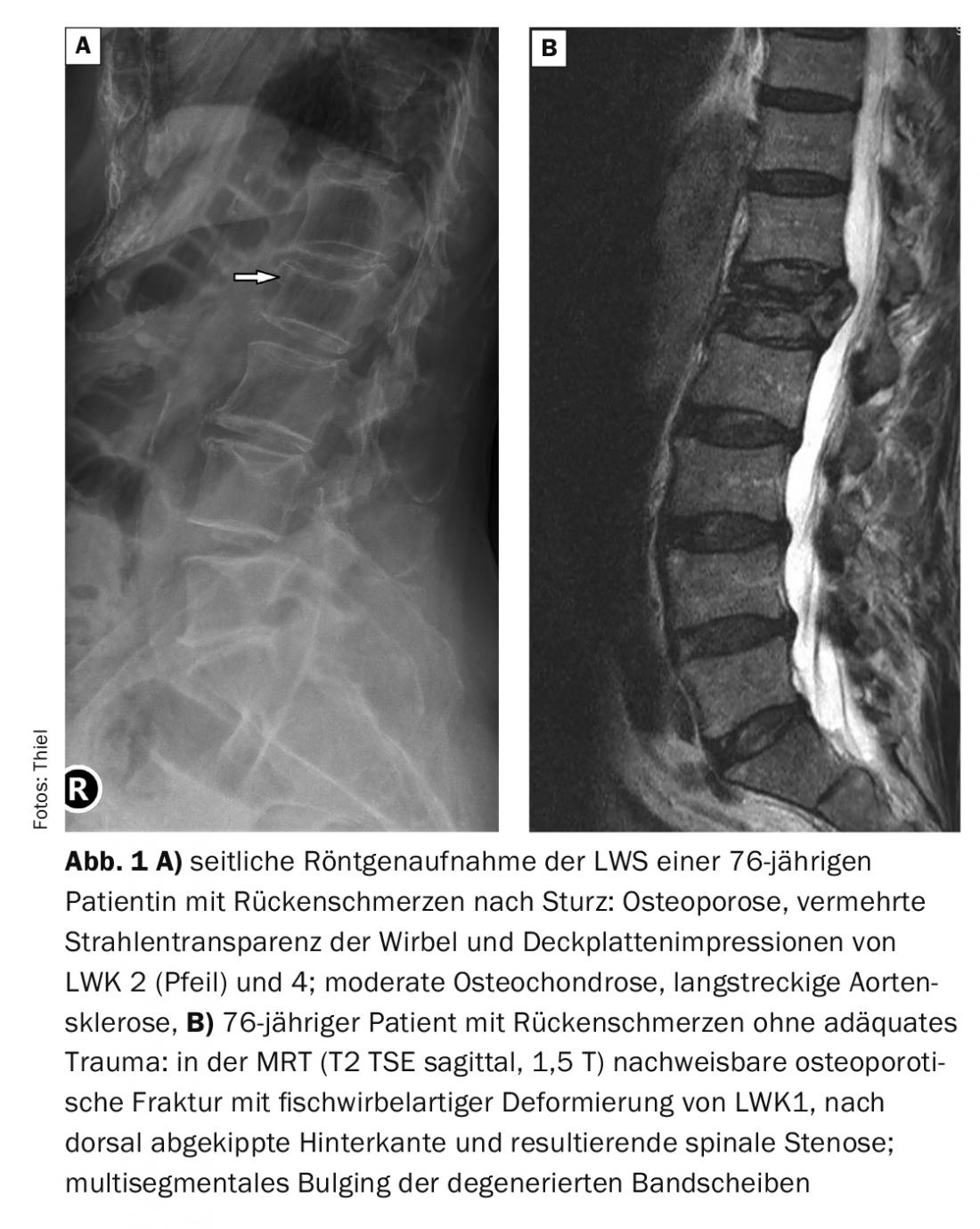
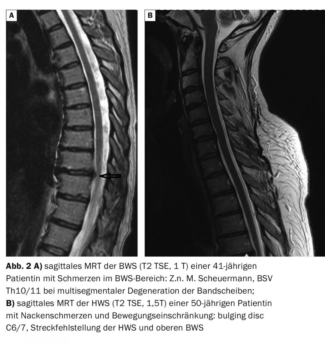
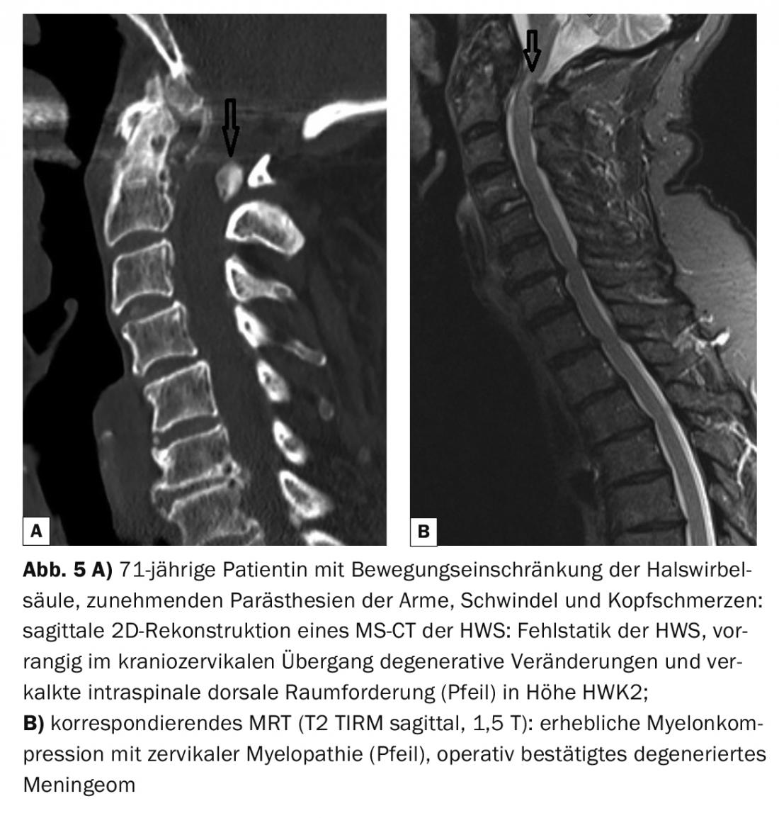
In the articles on the spine, various pathological changes are demonstrated:
- Traumatic (Fig. 1A, 1B)
- Degenerative (Fig. 2A, 2B)
- Inflammatory (Fig. 3A, 3B)
- Anomalies, as far as responsible for symptomatology (Fig. 4A, 4B).
- Tumorous (Fig. 5A, 5B)
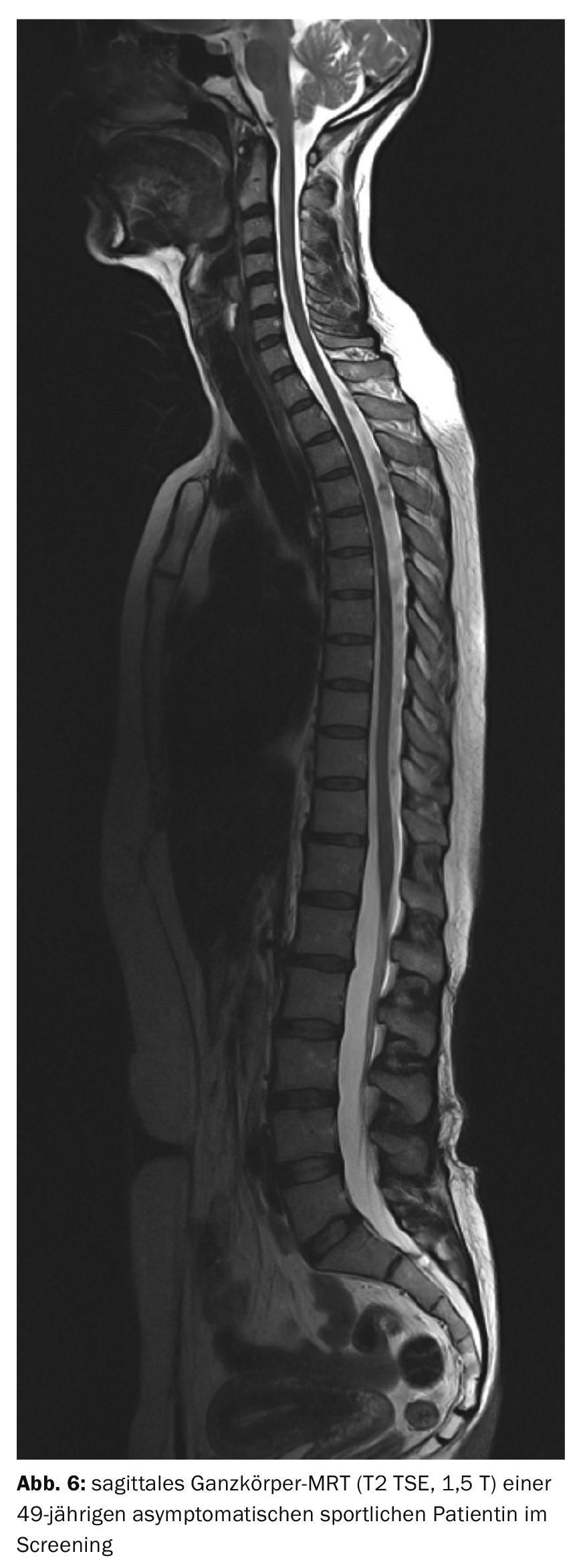
Among spinal imaging modalities, computed tomography and magnetic resonance imaging with assessment of complex anatomy are prominent in the value of evaluation. With the lack of radiation exposure of MRI, the procedure can be used especially in children and adolescents as well as in gravidity after the first trimester.The multiplanar imaging capability of pathologic processes, high soft tissue contrast, and also MR myelography as a noninvasive procedure make magnetic resonance imaging a valuable diagnostic tool. This also allows several spinal segments to be imaged together (Fig. 6) . The age-dependent different representation of the spinal structures in the findings must be taken into account [4].
In particular, the assessability of pathologic medullary processes is a domain of MRI. This is often associated with intravenous injection of gadolinium-containing contrast agents. A prerequisite for accurate image interpretation and reporting is the correct neurological classification of the clinical findings, knowledge of the medical history and a clearly defined question [1,3].

In most cases, computed tomographic examination is limited to a specific region of the spine, with the exception of whole-body multislice CT as part of staging in plasmacytoma patients. Axial scans and secondary multiplanar reconstruction (MPR) now also provide appropriate resolution of anatomic structures and pathologic changes, but soft tissue diagnosis is limited compared with MRI. In contrast to MR myelography, CT myelography requires intradural application of contrast agent and is therefore an invasive procedure. Frequent questions in CT are the extent of degenerative changes, traumatic lesions or anomalies of the skeleton due to anatomy and development. Great advantage of the procedure is the short examination time in trauma patients to diagnose bony injuries, possible myelon compression or bleeding.
X-rays of the spinal segments in two planes are still considered basic diagnostics today, but with limited informative value with regard to the exact disc situation and also intraspinal changes. Functional changes with segmental blockage or instability can be worked out by functional imaging. Anomalies of the vertebrae and variant rib systems can also be demonstrated radiographically [5]. Classic examples are the cervical ribs at the 7th cervical vertebra (consecutive nerve and/or vessel compression) or lumbosacral transitional vertebrae.
Summary
The spine is the localization of a variety of different changes and diseases. Anamnesis, clinical examination and laboratory findings lead to tentative diagnoses, which are clarified and differentiated by image morphology. The diseases affect all age groups and both sexes. They are a significant cost factor in the health care system due to diagnostic and therapeutic measures and place a considerable burden on national economies due to persistent incapacity to work and early retirement.
Literature:
- Stoller WD: Magnetic Resonance Imaging in Orthopaedics and Sports Medicine. Volume One. Third edition. Baltimore: Lippincott Williams & Wilkins 2007: 6-13.
- Thiel HJ: Cross-sectional diagnosis of the spine. MTA Dialog 7; 2008, 9: 554-557.
- Uhlenbrock D, ed: MRI of the spine and spinal canal. Stuttgart, New York: Georg Thieme Verlag 2001: 30.
- Vahlensieck M, Reiser M, eds: MRI of the musculoskeletal system. 2nd, completely revised and expanded edition. Stuttgart, New York: Georg Thieme Verlag 2002: 28-38.
- Wise R: Seventh cervical rib associated with subclavian artery occlusion and multiple infarcts: case report. J Neurosci Nurs 2008; 40(3): 169-172.
FAMILY PRACTICE 2019; 14(10): 38-39