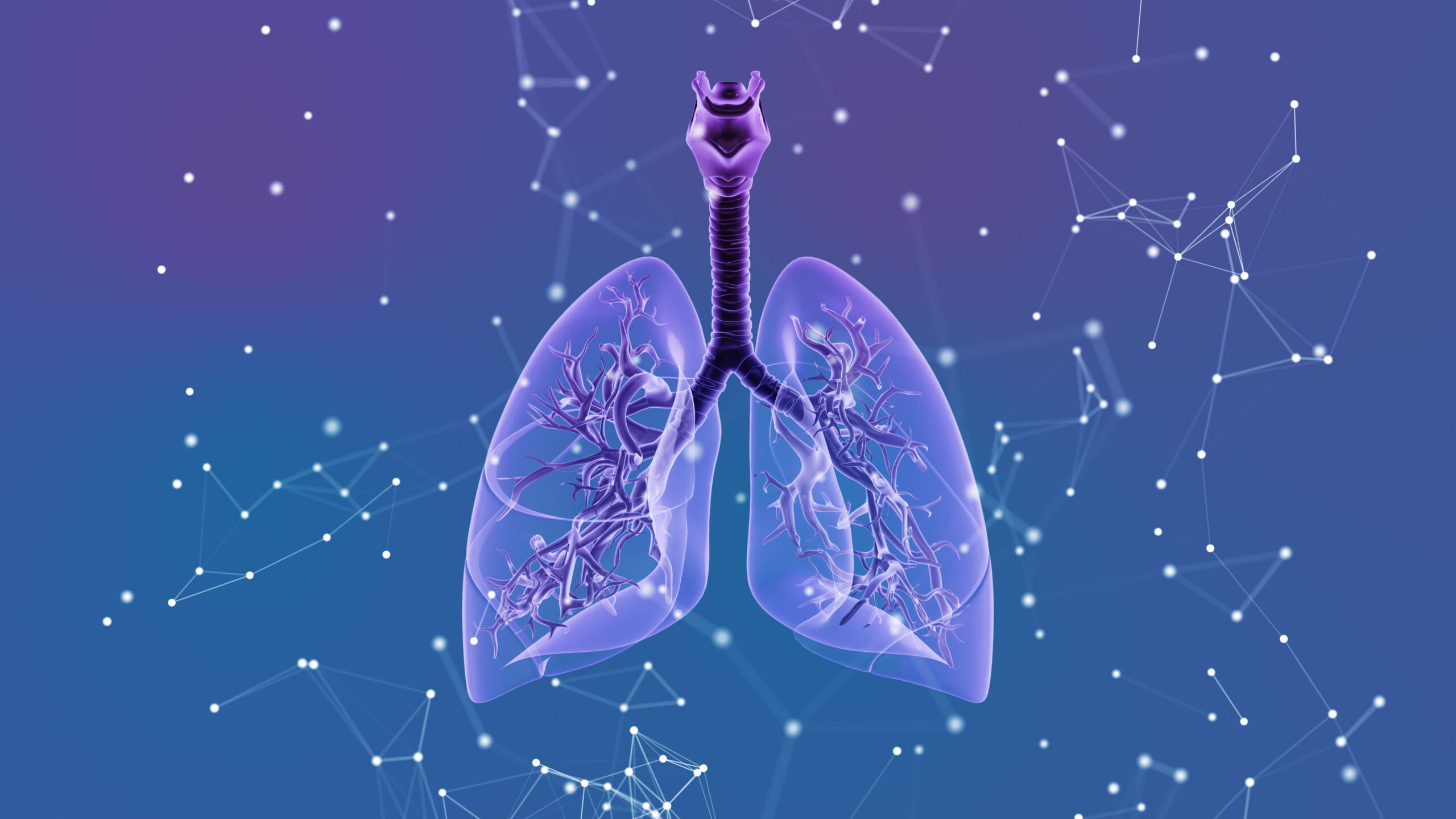
The formation of nephroliths is complex and dependent on many factors, which vary depending on the composition of the concrement and are still not fully understood. Many nephroliths remain asymptomatic and are discovered by chance during diagnostic imaging. One of the techniques that can be used to remove nephroliths is shock wave lithotripsy (ESWL), but there are certain complication risks to consider.
In a previous article, nephrolithiasis was reported as a possible cause of abdominal pain [2,4]. As a quick reminder, the incidence in Europe and the United States of America is about 0.5% per year, and the lifetime risk of disease is about 10-15%. The incidence of recurrence is approximately 50%, with a frequency of three or more recurrences occurring in 10-20% of stone carriers. About 80% of urinary stones are calcified. Nephrolithiasis is not just a problem of modern society. Kidney stones were already found in a 7000-year-old Egyptian mummy.
In the diagnosis of calculi, sonography and native computed tomography are useful. The stone discharge may cause colicky pain, a strong urge to urinate, and hematuria. By fitting a double-J splint with its ends in the renal pelvis and urinary bladder, urinary retention due to an occluding calculus can be avoided.
Extracorporeal shock wave lithotripsy (ESWL) can be used to break up kidney stones. The recurrence rate can be reduced by urinary alkalinization with medication, and it is also very important to drink sufficient amounts on a permanent basis.
ESWL is a therapeutic procedure in urology that allows the disintegration of urinary calculi by shock waves without anesthesia [4]. The method was developed in Munich and the first description was made by Chaussy in 1981. By the end of the last century, more than 150,000 patients worldwide had already been successfully treated with the method [1].
Kidney stones up to 2.5 cm in diameter can be treated with this method, as well as calculi in the upper ureter. Several smaller calculi up to a total stone volume of 5 cm³ can also be treated in this way [3]. Overview 1 provides information on the contraindications of ESWL.

According to the mechanism of shock wave generation, electromagnetic, electrohydraulic and piezoelectric ESWL are distinguished. The ESWL requirements listed in Overview 2 must be met.

Overview 3 and 4 provide information on the aftercare of ESWL and possible complications.


Case study
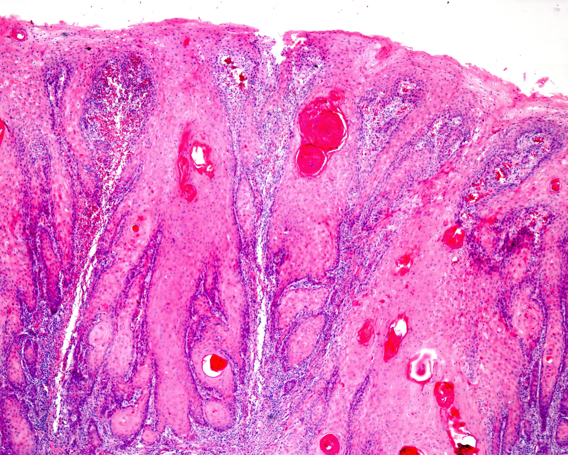
The case report (Figs. 1A to 1F) documents the therapeutic course of a patient who was 56 years old at the last CT scan and had bilateral colic with nephrolithiasis. Primary was a left DJ splint fitting in May 2022. With increasing shift of colic to the right side, a right ESWL was performed in January 2023. However, in addition to stone fragmentation, a complication had occurred with marked hemorrhage of the right kidney, resulting in a subcapsular hematoma with compression of the parenchyma in the upper half of the kidney, and marked hematuria, as documented by CT scans. Right flank pain required consistent analgesic treatment. During follow-up in early March, partial lysis of the renal hematoma was documented.

Take-Home Messages
- Urolithiasis is one of the common diseases in the field of urology.
- The symptomatology is broad.
- In addition to drug and minimally invasive treatment, extracorporeal shock wave lithotripsy has been established as a non-surgical procedure for a good 30 years.
- It is necessary to take into account some contraindications.
- Possible complications of therapy include renal hemorrhage.
Literature:
- Brendel W: Shock wave lithotripsy in kidney and gallbladder. In: Miehlke K.(eds) Verhandlungen der Deutschen Gesellschaft für Innere Medizin 1986; Vol 92. Munich: J.F. Bergmann-Verlag.
- Nephrolithiasis, https://flexikon.doccheck.com/de/Nephrolithiasis,(last accessed 08 Aug 2023).
- Extracorporeal shock wave lithotripsy (ESWL), www.uniklinikum-leipzig.de/einrichtungen/urologie/Seiten/behandlungsmethoden-eswl.aspx,(last accessed 08.08.2023).
- Manski D: Urology Textbook, www.urologielehrbuch.de/eswl.html,(last accessed 08.08.2023).
HAUSARZT PRAXIS 2023; 18(9): 40-42