As shown in this comparative study, the detection rate of the EUROArray Dermatomycosis Test is over 80% and can be increased up to 95% by combining it with direct microscopy. The test results are available very quickly (<24h).
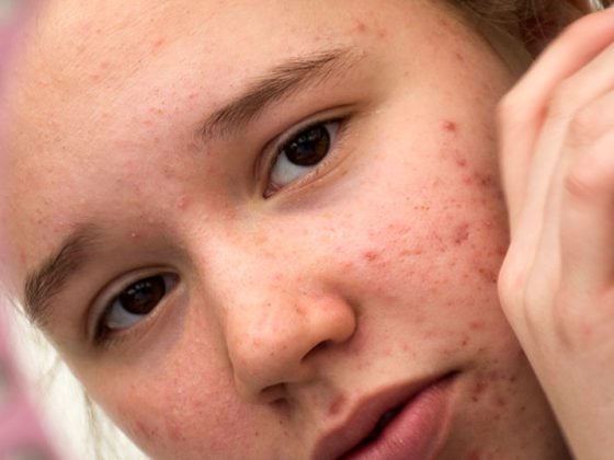
Mucosal skin infections are among the most common infectious diseases caused by dermatophytes, yeasts or molds. Up to 20% of the population of Western countries are affected. If mycosis is suspected, a laboratory diagnostic workup is required; however, the standard laboratory diagnostic procedure used to date is a relatively laborious workup procedure (direct microscopy followed by cellular culture and histologic examination).
Comparative study
The EUROArray Dermatomycosis method developed by EUROImmun AG in Lübeck, Germany, aims to optimize quality and efficiency through rapid, accurate and reliable diagnosis. It is a PCR-based microarray test system for the detection of all relevant dermatomycosis pathogens in one reaction (detection of all dermatophytes, assignment of the 23 species of dermatophytes and of 6 yeasts or molds). In a study by a research group at the University Hospital Schleswig-Holstein (D), a comparison was made with previous standard methods: direct microscopy (M) vs. cell culture (C) vs. PCR microarray analysis (P). The aim of the study was to identify the most reliable and time-saving method. Nail and skin specimen samples were recruited at Lübeck University Hospital and assigned to the patient group (n=140; 67 nail, 73 skin) or the control group (n=43; 24 nail, 19 skin) on the basis of visual examination. Each sample was analyzed using the three methods mentioned above.
EUROArray Dermatomycosis: high sensitivity and specificity
In 57% of the samples, mucosal infection could be confirmed by at least one of the three methods; in 43% of the cases, all three methods led to negative results. PCR microarray analysis achieved the highest detection rate (83.%–90,2%), followed by microscopy (39.0-66.6%) and cell culture (25.0-36%–36,6%6). A detection rate of 100% was achieved by using all three methods (M/C/P). When two different methods were combined, PCR microarray plus direct microscopy (M/P) resulted in the highest detection rate (94.4-95.1%), and classical diagnostics (M/C, 56.%–75,0%) achieved the lowest hit rate (70%). 8% of samples tested positive by cell culture were misdiagnosed.
Conclusion
The EUROArray Dernatomycosis is a test system that is simple to use and outperforms previous diagnostic methods in terms of sensitivity and time to diagnosis (<18 h). It also allows unambiguous species identification without the need for specific expertise and is therefore well suited for everyday clinical use. In cases of very exotic pathogens, which are not detected by the EUROArray Dermatomycosis Test System, the analytical sensitivity can be increased by additional direct microscopy. Using this combined test methodology, a detection rate of approximately 95% can be achieved and a rapid diagnosis is still possible (within less than 24 h). Identification by cell culture is only necessary in special cases, for example when the results are not compatible with the clinical picture.
Source: ADF, Munich (D)
Literature:
- Bieber K, et al: Munich Garching. Association for Dermatological Research. Poster presentation: P166 Fast and reliable diagnostics of fungal skin infections Annual Meeting ADF, March 2019.
Further reading:
- www.euroimmun.com/products/indications/molekulare-diagnostik/molekulare-infektionsdiagnostik/dermatomycosis.html
DERMATOLOGIE PRAXIS 2019; 29(3): 44