In chronic back pain, structural as well as functional changes in the brain are reported, which are related to the duration of pain. Neurological signatures identified using modern imaging are likely to play a supporting role in the prevention of chronic back pain in the future.
Why do some people often suffer from back pain even though there appears to be no identifiable injury or disease? This non-specific low back pain, mostly pain in the lumbar region and often also in the buttocks, is the most common form of back pain: In 85% of cases, no precise and pathoanatomically based diagnosis can be made [1]. It is important to note that the presence of non-specific back pain does not mean that it is pain of psychosomatic origin, but only that serious physical illness can be excluded as a cause. Understandably, many patients and clinicians are dissatisfied with the diagnosis of “nonspecific back pain,” with the common belief that back pain can be treated specifically depending on its presumed origin (e.g., originating from the disc, facet joint, or sacroiliac joint). Although treatment often helps, to date there is no clear evidence that “classification” based according to origin significantly increases treatment success [2]. In addition, symptoms of acute back pain usually improve spontaneously, with or without treatment. However, in 10-15% of patients the problem becomes chronic. One speaks of chronic back pain when the pain lasts longer than three months. Most treatment guidelines recommend patient pain education, physical activity and physical therapy, pharmacologic intervention with nonsteroidal anti-inflammatory drugs and opioids (for a short period of time), and spinal manipulation (e.g., chiropractic) for chronic low back pain [3]. The success rate of invasive procedures (e.g., spondylodesis) for chronic low back pain is usually no better than conservative treatments [4].
The health care costs of back pain are also worth noting: According to a study by the Swiss Federal Office of Public Health (2011), musculoskeletal disorders rank first in terms of indirect costs at with over 12 billion Swiss francs per year, of which around 7.5 billion are incurred for chronic back pain. A recent international health conference defined back pain as the greatest ailment worldwide, as measured by “years of living with a disability” [5]. These facts point to an urgent need for research to better understand chronic back pain, particularly the transition from acute to chronic and its causes, so that more effective prevention programs and therapies can ultimately be offered.
No pain without brain
Due to the often non-specific causes, a possible involvement of the brain in the pathogenesis of back pain is suggested in addition to unknown peripheral factors. Human pain perception is a complex phenomenon and the result of central nervous processing. Without the brain and its complex mechanisms of pain processing and modulation, subjective pain perception would not be possible, because the brain is not simply a passive receiver of nociceptive information. Nevertheless, the postulate of the French scientist and philosopher René Descartes, according to which a prick in the finger always triggers an identical pain reaction in the brain, is still stubbornly held by the almost 400-year-old and now obsolete postulate. Moreover, if the cause of the pain sensation is unknown, Descart’s dichotomy of body and soul is still frequently invoked, with suspicion being directed to an imagined or simulated pain. Today we know that pain can be felt without the presence of a nociceptive source – but the opposite can also occur. Furthermore, there is increasing evidence that chronic (back) pain is not only related to possible peripheral factors but also to changes in the brain. But how? Are these changes relevant in relation to pain or merely a side effect?
Brain changes in chronic pain
In the following, changes in the brain are divided into structural and functional changes, knowing that this division cannot be considered strictly dichotomous and that, for example, functional changes can accompany structural changes.
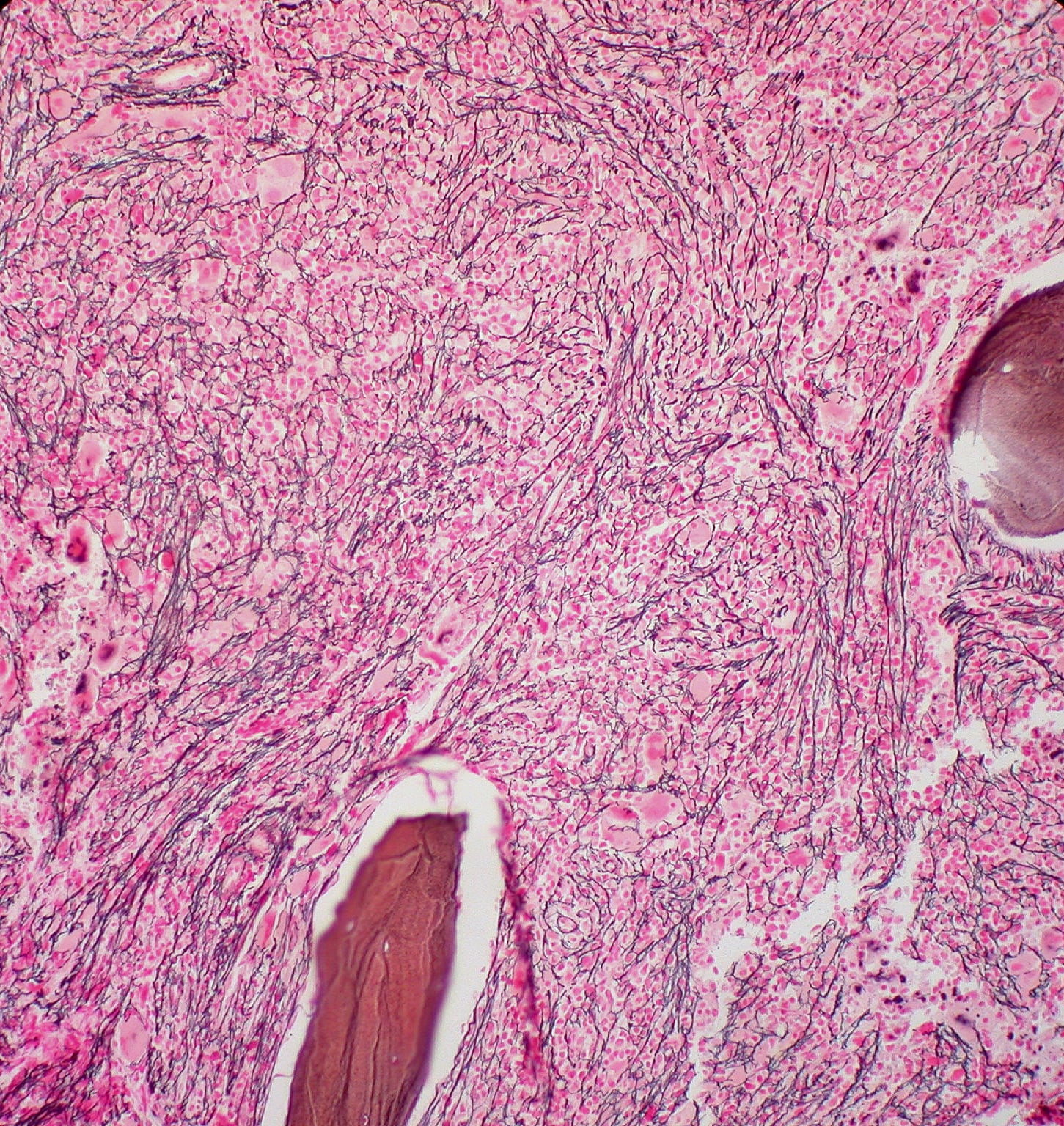
Structural changes: In recent years, a large number of studies have shown that structural brain changes in the form of gray and white matter volume changes occur in chronic pain patients. In the following, this article focuses on gray matter changes, as there is significantly more scientific evidence in this regard. Compared with healthy controls, chronic pain patients exhibit gray matter changes in quite a few brain regions, frequently reported changes in the dorsolateral prefrontal cortex, thalamus, brainstem, insula, primary somatosensory cortex (S1), and parietal cortex ( Fig. 1). However, the consensus among researchers ends with this observation. Much remains unclear and speculative.

A study from the University of Hamburg [6] investigated two important questions in this context: 1. Are these observed changes reversible and 2. Are these changes a consequence or cause of chronic pain? To answer these questions, the authors used voxel-based morphometry of magnetic resonance imaging data of the brain (structural brain scans), a commonly used method to study gray matter thickness/density. For this purpose, patients (n=32) with hip osteoarthritis were studied, a complaint with a clear peripheral cause that usually improves after joint replacement (88% of patients are pain-free after joint replacement) [7]. The results confirmed the findings on neuroplasticity of other chronic pain syndromes: Compared with a control group, patients showed significant changes in gray matter in a variety of brain regions. Another group of patients (n=10) was examined again 16-18 weeks after hip replacement surgery and these brain scans were compared with the preoperative brain scans. Here, the researchers discovered an increase in gray matter postoperatively in brain regions that showed atrophy preoperatively. This suggests that some of the observed brain changes are reversible, but this did not affect all brain regions studied. In addition, a comparison of the postoperative patient group with the healthy controls would have been necessary to show that there were no more changes between these groups in gray matter density. However, the authors reasonably conclude that the changes in gray matter are a consequence of chronic pain and not the cause. However, caution is advised here because, as the authors correctly discuss, it is probably not only the perception of pain that changes in patients when the “peripheral nociceptive generator” is removed, but also the lifestyle with social and sporting activities. Thus, it cannot be conclusively argued that the reversal of gray matter changes is a consequence of pain reduction. Nevertheless, based on these and similar studies, there is increasing evidence that these changes are reversible with effective treatment. Such changes in gray matter are also reported in pain patterns without a clear nociceptive source, for example, in phantom limb pain, fibromyalgia, migraine, or complex regional pain syndrome. For example, in a group of patients with different chronic pain patterns, it was shown that cognitive behavioral therapy over eleven weeks resulted in an increase in gray matter in different brain regions (prefrontal cortex, cingulate, parietal and somatosensory cortex). These changes were closely related to the success of the therapy, especially to the reduction of pain and its significance (catastrophizing) [8]. Moreover, in chronic back pain patients, the so-called sensory discrimination training, in which specific tactile and sensorimotor exercises are performed, seems to have positive effects on neuroplasticity and pain perception [9].
Functional changes: Four years ago, a team of researchers at the University of Chicago published fascinating results [10]. Using functional magnetic resonance imaging (fMRI), the researchers compared the brain activity of patients with a back pain duration of approximately two months (acute/sub-acute, n=94) with patients who suffered from back pain for more than ten years (chronic, n=59). Acute pain always involves a certain pattern of brain activity (also known as the pain matrix or neurological signature of pain). Now the patients with acute back pain showed that well-known activity pattern associated with acute pain. In comparison, however, chronic pain patients showed an altered activity pattern: Acute pain brain regions showed less neuronal activity, whereas significantly more activity was observed in emotion-associated regions (prefrontal cortex, amygdala). The authors also demonstrated the same effect in a longitudinal study design: Pain-related brain activity changed toward emotion-associated activity in those patients who became chronic, but not in those who recovered after the acute phase. The study was thus able to show concretely for the first time that back pain in the acute and chronic stages has different brain activity patterns. In addition, a changing neurological signature could serve as a biomarker for identifying individuals who are prone to chronicity (but it must be emphasized that this is currently still a pipe dream).
These results are supported by the knowledge that psychosocial factors play a major role in the chronification of back pain and that somatic factors are increasingly fading into the background. Of particular importance is the fear-avoidance model, which describes how unfavorable cognitive beliefs and perceptions can negatively influence disease development in exercise-related pain. Meanwhile, we can study and map such processes in the brain.
In the latest fMRI study of our research group, we examined 20 patients with chronic low back pain and varying degrees of fear-avoidance behavior (measured by well-validated questionnaires), focusing on the connectivity of two brain regions that are strongly implicated in supraspinal pain modulation (Fig. 2): The periaqueductal gray (PAG) and the amygdala. During the fMRI recordings, participants were presented with standardized videos (duration: 4 seconds) of potentially dangerous movements for the back in everyday life (e.g., vacuuming, picking up a heavy flowerpot). Neutral movements such as leisurely stair walking formed the control condition. Analysis showed that patients had significantly lower amygdala-PAG connectivity compared to healthy control subjects. However, this is only during the viewing of the potentially dangerous videos, not in the neutral condition. Also, the strength of this connectivity correlated negatively with the expression of the patients’ fear-avoidance behavior: The higher the score on the questionnaire, the lower the connectivity between the amygdala and the PAG. These are likely to be the first indications of how fear and avoidance behaviors, as a psychological component, influence biological systems of pain modulation, possibly contributing to the chronification of back pain.

Open questions and a look into the future
Many questions are still open: Are there specific changes in the brain depending on the chronic pain pattern or do these represent a general effect in any type of chronic pain? Are there changes that can be characterized as vulnerability factors for chronic pain?
The current evidence clearly suggests that the brain changes structurally as well as functionally in chronic back pain, but we do not yet know exactly what these changes mean. One of the authors of this article (PS) led a study suggesting that various mechanisms may lead to gray matter changes and that neurodegenerative processes are unlikely to be the cause [11]. Furthermore, most study results come from correlation analyses, which do not allow causal conclusions. Nevertheless, in the future, these neurological signatures could be useful in identifying subgroups of chronic back pain patients or even in determining risk factors. However, the potential effect of spinal manipulation on the brain in chronic back pain could also be quantified using modern imaging. In addition to other promising methods such as functional near-infrared spectroscopy [12], functional imaging methodology, most notably fMRI, continues to refine. Recent fMRI studies reveal changes in the network structure of the brain of chronic pain patients, with specific changes depending on the duration of pain [13]. Meanwhile, it is also possible to identify chronic back pain patients using only brain activity patterns and specific algorithms, this with a prediction accuracy of over 90% [14]. If these studies can be confirmed in several research laboratories around the world, new statements and questions could be generated on an individual level.
Take-Home Messages
- In up to 85% of patients with back pain, no precise diagnosis can be made, with the brain playing an important role in addition to unknown peripheral factors.
- In chronic back pain, structural as well as functional changes in the brain are reported, which are related to the duration of pain.
- These changes in the brain are reversible with appropriate therapy and correlate with pain reduction.
- Neurological signatures identified using modern imaging that are sensitive to the development and progression of back pain are likely to play a supporting role in the prevention of chronic back pain in the future.
Literature:
- Deyo RA, Weinstein JN: Low back pain. The New England journal of medicine 2001; 344: 363-370.
- Kamper SJ, et al: Treatment-based subgroups of low back pain: a guide to appraisal of research studies and a summary of current evidence. Best practice & research. Clinical rheumatology 2010; 24: 181-191.
- Dagenais S, Tricco AC, Haldeman S: Synthesis of recommendations for the assessment and management of low back pain from recent clinical practice guidelines. The spine journal: official journal of the North American Spine Society 2010; 10: 514-529.
- Maher C, Underwood M, Buchbinder R: Non-specific low back pain. Lancet (London, England) 2017; 389: 736-747.
- Hoy D, et al: The global burden of low back pain: estimates from the Global Burden of Disease 2010 study. Annals of the rheumatic diseases 2014; 73: 968-974.
- Rodriguez-Raecke R, et al: Brain gray matter decrease in chronic pain is the consequence and not the cause of pain. The Journal of neuroscience: the official journal of the Society for Neuroscience. 2009; 29: 13746-13750.
- Nikolajsen L, et al: Chronic pain following total hip arthroplasty: a nationwide questionnaire study. Acta anaesthesiologica Scandinavica 2006; 50: 495-500.
- Seminowicz DA, et al: Cognitive-behavioral therapy increases prefrontal cortex gray matter in patients with chronic pain. The journal of pain : official journal of the American Pain Society 2013; 14: 1573-1584.
- Kalin S, Rausch-Osthoff AK, Bauer CM: What is the effect of sensory discrimination training on chronic low back pain? A systematic review. BMC musculoskeletal disorders 2016; 17: 143.
- Hashmi JA, et al. Shape shifting pain: chronification of back pain shifts brain representation from nociceptive to emotional circuits. Brain: a journal of neurology 2013; 136: 2751-2768.
- Pomares FB, et al: Histological Underpinnings of Grey Matter Changes in Fibromyalgia Investigated Using Multimodal Brain Imaging. The Journal of neuroscience: the official journal of the Society for Neuroscience 2017; 37: 1090-1101.
- Vrana A, et al: Cortical Sensorimotor Processing of Painful Pressure in Patients with Chronic Lower Back Pain-An Optical Neuroimaging Study using fNIRS. Frontiers in human neuroscience 2016; 10: 578.
- Mansour A, et al: Global disruption of degree rank order: a hallmark of chronic pain. Scientific reports 2016; 6: 34853.
- Callan D, et al: A tool for classifying individuals with chronic back pain: using multivariate pattern analysis with functional magnetic resonance imaging data. PloS one (2014) 9: e98007.
- Cauda F, et al: Gray matter alterations in chronic pain: A network-oriented meta-analytic approach. Neuroimage Clin 2014; 4: 676-686.
- Meier ML, et al: The impact of pain-related fear on neural pathways of pain modulation in chronic low back pain. PAIN Reports 2017.
InFo NEUROLOGY & PSYCHIATRY 2017; 15(3): 4-8.