The standard of care for epithelial tumors is excision with a safety margin and surgical closure of the defect. In addition, there are local, radiotherapeutic and systemic approaches, depending on the tumor type and situation. Each patient can thus be offered the most appropriate therapy.
Epithelial tumors (“white skin cancer”) are the most common tumors in humans. Basal cell carcinoma (BCC) accounts for approximately 80% of all non-melanocytic skin tumors. The most common histologic type is the nodular form, ahead of the sclerodermiform and superficial types. Even if it hardly metastasizes (in less than 0.5% of all cases), BZK grows aggressively locally. Early detection and consistent therapy are therefore essential. The individual lifetime risk of suffering a BCC is 30% for Caucasians [1].
The second most common white skin cancer is spinal cell carcinoma (SCCHN). This often develops from actinic keratoses (precancerous lesions). In Europe, there are around 30 new cases per 100,000 people per year, but incidence rates are increasing rapidly (50-200% in the last 30 years [2,3]). SCCHN may rarely (in 2-5%) metastasize to regional lymph nodes and may also form distant metastases. In cases of distant metastasis, the median survival rate is less than two years.
Due to the rapid increase in skin cancer cases in Switzerland, those treating patients are increasingly confronted with the question of which form of therapy is most suitable for the individual patient. In this decision, the focus is always on the radicality of the tumor treatment, which spares the patient with basal cell carcinoma a recurrence that could lead to further treatment or even a destructive ulcerative tumor. In the case of SCLC, this can additionally reduce the risk of metastasis.
However, other aspects are important for the patient: The function of the affected body part must be preserved whenever possible. Since these tumors are most frequently located on the face, the aesthetic result after tumor removal also plays a major role for many of those affected.
Cutting
Simple excision is the best treatment option for a majority of epithelial tumors because it completely removes the tumor with relatively little effort in a single session. In addition, histologic workup can document the totality of tumor removal. In small, clinically typical tumors, this treatment can also be used simultaneously diagnostically and curatively without prior biopsy. Tumors should always be excised with a safety margin in healthy tissue, although there is no consensus in the literature on the size of the necessary safety margins.

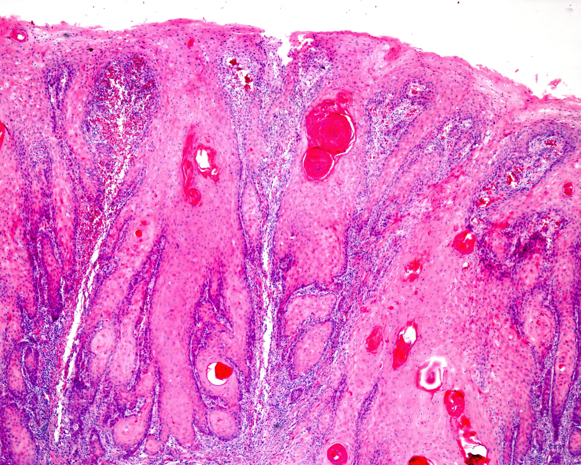
For basal cell carcinoma, the safety distance should generally be 3-5 mm. In nodular BCC (Fig. 1), this achieves recurrence rates of less than 5%. However, in infiltrative (cirrhotic) BCC (Fig. 2), larger safety margins are required to remove the tumors curatively. Clinical safety distances of 13-15 mm are necessary here for a recurrence rate below 5%.

Larger safety margins of 5-7 mm are also necessary for spinocellular carcinoma, depending on the histologic degree of differentiation.
The excisional defect can usually be closed directly in smaller tumors removed in this way after completion to a spindle-shaped defect. Because virtually all surgeries can be performed under local anesthesia with very little distress to the patient, there are almost never patients who are considered inoperable for skin cancer surgery. Concomitant medications such as anticoagulants do not usually need to be discontinued.
Mohs surgery (micrographically controlled surgery): Significantly lower recurrence rates of 1-2% with a smaller safety margin can be achieved with Mohs surgery. Tumors are excised primarily with a small safety margin of 1-2 mm. The excised specimens are then examined histologically after cryofixation. If tumor cells are still found in the incision margin, a re-excision can be directly followed and the same procedure repeated. What is important in this method is that the specimens are processed using a special technique so that the entire margin of the section can be assessed in contrast to normal cross-sectional histology, in which only a fraction of the margin is examined at a time and margin-forming tumor extensions between two cross-sections can be missed. In addition, with this method, the evaluation of the histological specimen must be performed by the dermatosurgeon himself, as this is the only way to perform a precise re-excision without unnecessary tissue loss.
Thus, recurrence rates of 1-2% can be achieved in primary basal cell carcinoma. For recurrent basal cell carcinoma, recurrence rates as high as 17% with ordinary excision can be reduced to less than 5% with Mohs surgery. Particularly when excisional defects become so large that reconstruction with flapplasty or skin grafting must be performed, it is important that tumor-free status be perfectly established prior to defect closure. In addition, Mohs surgery usually results in smaller excision defects due to the smaller safety margins, which in turn leads to less costly reconstructions and better esthetic results.
However, due to the more complex logistics and the close collaboration required between dermatosurgeons and histology laboratories, this method must be limited to tumors in the facial region that require reconstruction by flap surgery or full-thickness skin graft, tumors with infiltrative growth patterns that would necessitate a large safety margin (e.g., cirrhotic basal cell carcinoma), recurrent tumors, and patients with special risk factors such as immunosuppression (Tab. 1). For these tumors, treatment alternatives such as topical therapies are not an option in most cases. If the infrastructure for Mohs surgery is not available and in the presence of tumors that are difficult to evaluate histologically in frozen section (e.g., de-differentiated SCK), one can switch to a variant of section margin-controlled surgery with histologic examination on kerosene section. However, this is much more costly for the patient due to multiple surgical appointments. The advantage of the smallest possible excision defect due to the exact localization of the margin-forming tumor parts is lost.

Local treatments
Cryosurgery: In some situations, cryosurgery with liquid nitrogen (-196°) can be a good alternative to normal surgery. In particular, precancerous lesions and superficial tumors such as trunk skin basaliomas (superficial BCC) can be treated easily and with good cure rates. Anesthesia is usually not necessary. However, the postoperative result may be affected by hypopigmented scars, especially in dark skin types. In invasive tumors, cryosurgery is used only in exceptional cases when ordinary excision is not possible or is rejected by the patient. However, with freezing and thawing times adapted to tumor thickness, satisfactory results can still be achieved. The lack of histologic control results in higher recurrence rates.
Topical treatments: Topical treatments with creams are only approved and useful for precancerous lesions and the superficial form of BZK (sBZK). 5-Fluorouracil (5-FU), the cytostatically active antimetabolite and inhibitor of thymidylate synthase, is used in cream form twice daily for actinic keratoses, Bowen’s disease, and sBZK until ulceration occurs. Cure rates of approximately 80-86% have been demonstrated. Side effects include scarring.
Imiquimod-5% cream binds to Toll-like receptor 7, and this activation results in the release of proinflammatory cytokines and an inflammatory response. The cream is applied three times a week for actinic keratoses and over five days a week for sBZK. The histologic cure rate for sBZK is approximately 80% [4].
Photodynamic therapy (PDT): this can be performed with 5-aminolevulinic acid or its ester methyl-aminolevulinic acid for actinic keratoses, Bowen’s disease, and superficial BCC (treating the same region twice). Thin nodular BCCs can also be treated with PDT after curettage, but the results are less reliable and the treatment is off-label. In the meantime, the Daylight PDT variant also exists, in which daylight is used instead of the red light lamp. This results in a significant reduction in pain. PDT shows a very good cosmetic result, with hypopigmentation not occurring at all. It can also be repeated as often as desired, especially in immunosuppressed patients.
Radiotherapy
Surface radiation therapy is another useful alternative in the treatment of non-melanoma skin cancer (NMSC). It is especially considered when surgery is rejected, whether by patient request, extent of the finding, or general condition and concomitant diseases. Accordingly, this therapeutic modality is frequently used in elderly patients (over 60 years of age).
As a rule, generator voltages of 10-50 kV are used. The classic indications for curative treatment involve extensive areal precancerous lesions, Bowen’s disease, nodular basal cell carcinoma, and lentigo maligna, where excellent results are achieved.
Recurrence rates of 5% (well-differentiated solid form) to 31% (sclerodermiform growth forms) have been described for basal cell carcinoma in correlation with the underlying histology [5]. The 5-year recurrence rates for spinocellular carcinomas are approximately 10% (well-differentiated tumors) and approximately 23% (dedifferentiated forms) [6,7].
The advantages of radiotherapy are excellent cosmetic results (especially on the face) and painless as well as tissue-sparing treatment of extensive forms and cases at difficult anatomical localization. Disadvantages can be considered the required number of sessions, permanent hair loss and postactinic skin changes (atrophies, telangiectasias). Also, in case of a possible recurrence, further irradiations at the same site are only possible to a limited extent or not at all.
Acute side effects include erythema, tumor necrosis, and wound and crust formation with subsequent healing. As a rule, they are intentional and can be observed within the framework of radiobiological principles. Chronic effects include depigmentation and, as mentioned, skin atrophy and telangiectasias, which often take years to develop. The risk of malignancy induction is extremely low when dosing instructions are followed.
System therapy for locally advanced or metastatic basal cell carcinoma
For the past few years, the specific smoothened inhibitor vismodegib (and now sonidegib) has been approved for locally advanced or metastatic BCC for which surgical treatment or radiotherapy cannot be performed. The indication should be discussed on an interdisciplinary basis at a tumor center. Response rates of 76% for locally advanced BCC and 38% for metastatic tumors have been described for vismodegib [8]. Side effects are common and include muscle spasms, diffuse alopecia, taste disturbances, weight loss, and fatigue. Therefore, application pauses must often be carried out.
Literature:
- Guidelines Program Oncology, German Cancer Society DK, AWMF: S3 Guideline. Prevention of skin cancer. 2014.
- Lomas A, Leonardi-Bee J, Bath-Hextall F: A systematic review of worldwide incidence of nonmelanoma skin cancer. Br J Dermatol 2012; 166: 1069-1080.
- Stratigos A, et al: Diagnosis and treatment of invasive squamous cell carcinoma of the skin: European consensus-based interdisciplinary guideline. Eur J Cancer 2015 Sep; 51(14): 1989-2007.
- Arits AH, et al: Photodynamic therapy versus topical imiquimod versus topical fluorouracil for treatment of superficial basal-cell carcinoma: a single blind, non-inferiority, randomised controlled trial. Lancet Oncol 2013; 14: 647-654.
- Zagrodnik B, et al: Superficial radiotherapy for patients with basal cell carcinoma: recurrence rates, histologic subtypes, and expression of p53 and Bcl-2. Cancer 2003; 98: 2708-2714.
- Barysch MJ, et al: Long-Term Recurrence Rate of Large and difficult to treat cutaneous squamous cell carcinomas after superficial radiotherapy. Dermatology 2012; 224: 59-65.
- Panizzon RG, Dummer R, Beyeler M: Radiotherapy of malignant skin tumors. Physical therapy interventions in dermatology 2003; 135-139.
- Basset-Seguin N, et al: Vismodegib in patients with advanced basal cell carcinoma (STEVIE): a pre-planned interim analysis of an international, open-label trial. Lancet Oncol 2015; 16: 729-736.
DERMATOLOGIE PRAXIS 2017; 27(2): 15-18