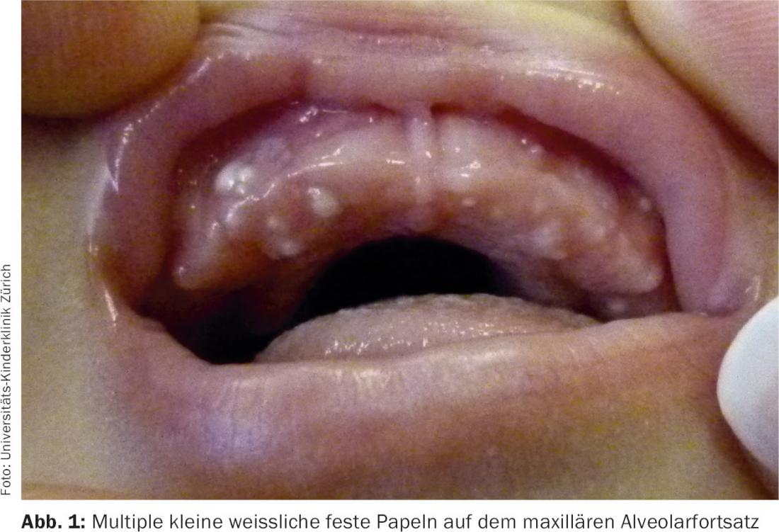
Case report: At the two-month check-up, a healthy female infant shows multiple small whitish nodules on the maxillary gingiva. The child was drinking well and did not appear to be bothered in any way by the mucosal changes. The question arises as to the genesis and therapeutic necessity of the nodules.
Clinical picture: 2-month-old infant in very good general condition with multiple pale, firm small nodules along the upper dental arch (Fig. 1). The rest of the infant’s examination is unremarkable.
Quiz
Based on this information, what is the most likely diagnosis?
A Epstein Beads
B Gingival papillomas
C vesicles in herpes simplex initial manifestation.
D Gingival cysts
E Congenital epulis
Correct answer and diagnosis: The correct answer is D. These are gingival cysts.
Diagnosis and Discussion:
The clinical picture, the patient’s age, and the good concomitant general condition are typical for gingival cysts.
- Enoral epithelial cysts in small infants were first classified by Fromm in 1967 according to histopathologic criteria and the location of the cysts. Fromm distinguished between Epstein’s beads, Bohn’s nodules, and inclusion cysts [1].
- Epstein beads can typically be found on the raphe mediana of the hard palate and correspond to epithelial tissue entrapped during fusion of the raphe.
- Bohn’s nodules are usually located on the lingual and, less commonly, vestibular surface of the maxillary gingiva or are located on the anterior hard palate. While Fromm assumed that these cysts contain mainly mucous gland remnants and excretory ducts, other authors assume an exit from the dentallamina when they are located at the alveolar process [2].
- Dental inclusion cysts , on the other hand, are mainly located on the alveolar process in the form of one to numerous small whitish solid papules ranging in size from 1 to a maximum of 3 mm. They arise from epithelial remnants of the dentallamina and histopathologically show concentrically layered horn lamellae.
Due to the frequent confusion of terms and also the difficult clinical differentiation of Bohn’s nodules and dentallamina cysts in particular, a new classification was already introduced by Jorgenson in 1982, in which only palatal and alveolar or gingival cysts are now differentiated depending on the localization [2].
Enoral cysts are most commonly present congenitally, but more rarely are described to occur in the neonatal period. A 2012 study by Monteagudo et al. published study [3] showed a prevalence of 53.7% for palatal cysts and 13.4% for gingival cysts in 1000 newborns examined. Both palatal and gingival cysts were found in 11% of the children. In the study, there was also a girl turnout and an increased incidence in mature and transferred infants. These figures are well compatible with other studies where palatal cysts were found in 55-86% of all newborns and gingival cysts in 13.8-97% [3,4].
In general, no therapeutic intervention was necessary in these various studies. The cysts usually regressed within the first 6 months of life [5].
Gingival papillomas are extremely rare in the neonatal period. They present in adolescence and adulthood as skin-colored, usually rather soft enoral papules on the gingiva, tongue and palate and are seen either as an expression of HPV infection or also in the context of syndromal diseases such as Cowden syndrome. Infection with herpes simplex virus is likely to cause pain and, in infancy, often increased body temperature. In addition, the primary florescence in this case is a vesicle and not a solid papule. In congenital epulis, an isolated swelling is seen rather than multiple small firm nodules, as in this case.
Abstract: Enoral epithelial cysts are very common according to the current literature and can therefore be considered as normal variants. While Epstein pearls – palatal cysts along the raphe mediana – are well known to many medical colleagues, the existence of gingival cysts is somewhat less familiar. Such a case in pronounced forms can therefore lead to a certain uncertainty of the parents and also of the treating physicians.
Knowledge of the various enoral cysts helps reassure the parents of the affected child and avoid unnecessary and often painful diagnostic procedures.
Literature:
- Fromm A: Epstein’s pearls, Bohn’s nodules and inclusion-cysts of the oral cavity. J Dent Child 1967; 34: 275-287.
- Jorgenson RJ, et al: Intraoral findings and anomalies in neonates. Pediatrics 1982; 69: 577-582.
- Monteagudo B, et al: Prevalence of Milia and Palatal and Gingival Cysts in Spanish Newborns. Pediatric Dermatology 2012; 29: 301-305.
- Paula JD, et al: Oral and facial inclusion cysts in newborns. J Clin Pediatr Dent 2006; 31: 127-129.
- Van Heerden WFP, et al: Diagnosis and management of oral lesions and conditions in the newborn. South African Family Practice 2010; 52: 6, 489-491.
DERMATOLOGIE PRAXIS 2016; 26(6): 42-43