Pseudoxanthoma elasticum (PXE), also known as Grönblad-Strandberg syndrome, is a rare hereditary disease that primarily affects the skin, eyes and arterial vascular system. Early diagnosis is crucial for the timely treatment of extracutaneous complications.
PXE is caused by mutations in the ATP-binding cassette transporter protein 6 (ABCC6). The estimated prevalence worldwide is between 1:25,000 and 1:70,000, although a large number of undetected cases are suspected due to the difficulty of diagnosis [1–4]. In the early stages, the skin usually shows pronounced lesions, with extradermal manifestations occurring later [1]. Darier first named the skin disease “Pseudoxanthoma elasticum” in 1896 [5]. The Swedish ophthalmologist Ester Grönblad and the dermatologist James Strandberg discovered a connection between skin and ocular fundus changes in 1926, which is why the disease is also known under the synonym Grönblad-Strandberg syndrome [6].
| PXE is a rare disease whose early diagnosis is clinically relevant as it can be associated with cardiovascular and retinal manifestations. The diagnosis is based firstly on the histopathological findings and secondly on the detection of a mutation in the ABCC6 gene. The earlier the diagnosis is made, the sooner preventive measures and close monitoring can be taken, which can have a significant impact on the prognosis. |
| according to [1] |
Predilection sites for the skin changes are the neck, the elbow bends, the trunk and the periumbilical region. Diagnosis is based on clinical examination, histological analysis of the lesions and, if necessary, molecular genetic testing to detect the gene mutation. Histopathology shows clumping and mineralization of the elastic fibers and fragmentation of the elastic fibers in the middle/deep dermis [2,3]. Cardiovascular manifestations such as hypertension, angina pectoris and intermittent claudication are among the main causes of morbidity in PXE patients. Early atherosclerosis may develop due to mineralization of the inner elastic lamina of the blood vessels and a lipid change with a reduction of the HDL cholesterol level in the blood plasma as well as hypertriglyceridemia.

12-year-old girl with papular changes
The patient of Caucasian origin presented in November 2016 because of sporadic, painless, slightly itchy yellow papules (xanthoma) in the neck region that had been developing for around a year [1]. The lesions have not yet been treated. The child had no pain or signs of inflammation in the lesions or surrounding areas and there were no associated symptoms. The patient was otherwise healthy, without regular medication and had no relevant personal or family history of dermatoses.
Inspection: Painless, uneven, rough and yellow plaques without signs of inflammation were found in the posterior neck region, which spread bilaterally and symmetrically to the right and left neck region. They gave the skin a parchment-like appearance. The rest of the integument showed no changes.
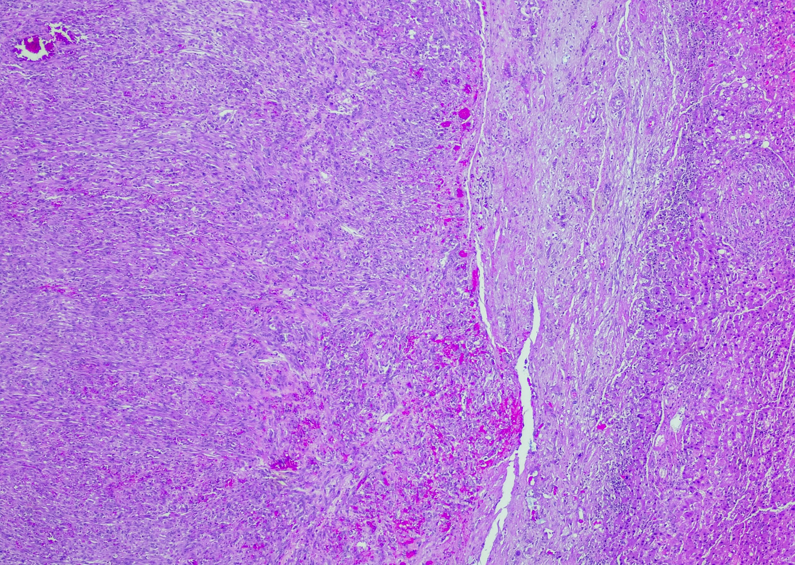
Skin biopsy: The results of the skin biopsy revealed clumped and fragmented fibers mainly in the middle dermis and beyond in the core of the elastic fibers. Von Kossa staining can be used to demonstrate that these are predominantly calcium-phosphate deposits [7]. There was no evidence of mineralization or mucin deposition. It is not yet clear whether the calcification of the elastic fibers is secondary to the fragmentation or whether the fragmentation is the primary cause of the calcification.
Further examinations: Blood count with lipid profile and urinary sediment were unremarkable. The ECG, echocardiogram, ultrasound of the carotid artery and the aortoiliac veins and arteries were also normal, and the ophthalmologic examination revealed no abnormal findings.
Diagnosis and course: The pathology specialist decided that ABCC6 gene testing was not necessary due to the clinical aspect of the lesions, which were very characteristic of PXE. Since then, the patient has been examined annually by ophthalmologists, cardiologists and dermatologists. As of February 2019, no systemic manifestations have been reported.
ABCC6 gene mutation
In 2000, mutations in the ABCC6 gene were identified as the cause of PXE. To date, over 300 different mutations in the ABCC6 gene are known in PXE patients. The most frequently occurring mutations are the nonsense mutation p.R1141X (c.3421C>T, rs72653706) in exon 24 and a deletion of exons 23 to 29 (c.2996_4208del) on at least one allele [8].
Literature:
- Lucas C, et al: Case Report: Pseudoxanthoma elasticum. F1000Res. 2020 Jan 9; 9: 9. doi: 10.12688/f1000research.21431.1.
- Marconi B, et al: Pseudoxanthoma elasticum and skin: Clinical manifestations, histopathology, pathomechanism, perspectives of treatment. Intractable Rare Dis Res 2015; 4(3): 113-122.
- Lopes L, et al: Elastólise da derme papilar semelhante a pseudoxantoma elástico. Rev Soc Port Dermatol Venereol 2014; 72: 223-226.
- Plomp, AS, et al: Proposal for updating the pseudoxanthoma elasticum classification system and a review of the clinical findings. American Journal of Medical Genetics 2010; Part A 152: 1049-1058.
- Grimmer H: [Histological iconography. Pseudoxanthoma elasticum (Darier 1896)]. Z Skin Sex cr. 1961 Apr 1; 30: xxvii-xxx. German. PMID: 13708725.
- Groenblad E: Angioid streaks-pseudoxanthoma elasticum. Acta Ophthalmologica 1929; 7: 329-329.
- Otkjaer-Nielsen A, et al: Apatite crystals in pseudoxanthoma elasticum: a combined study using electron microscopy and selected area diffraction analysis. JID 1977; 69: 376-378.
- Pfendner EG, et al: Mutation detection in the ABCC6 gene and genotype-phenotype analysis in a large international case series affected by pseudoxanthoma elasticum. Journal of medical genetics 2007; 44: 621-628.
DERMATOLOGY PRACTICE 2024; 34(2): 46