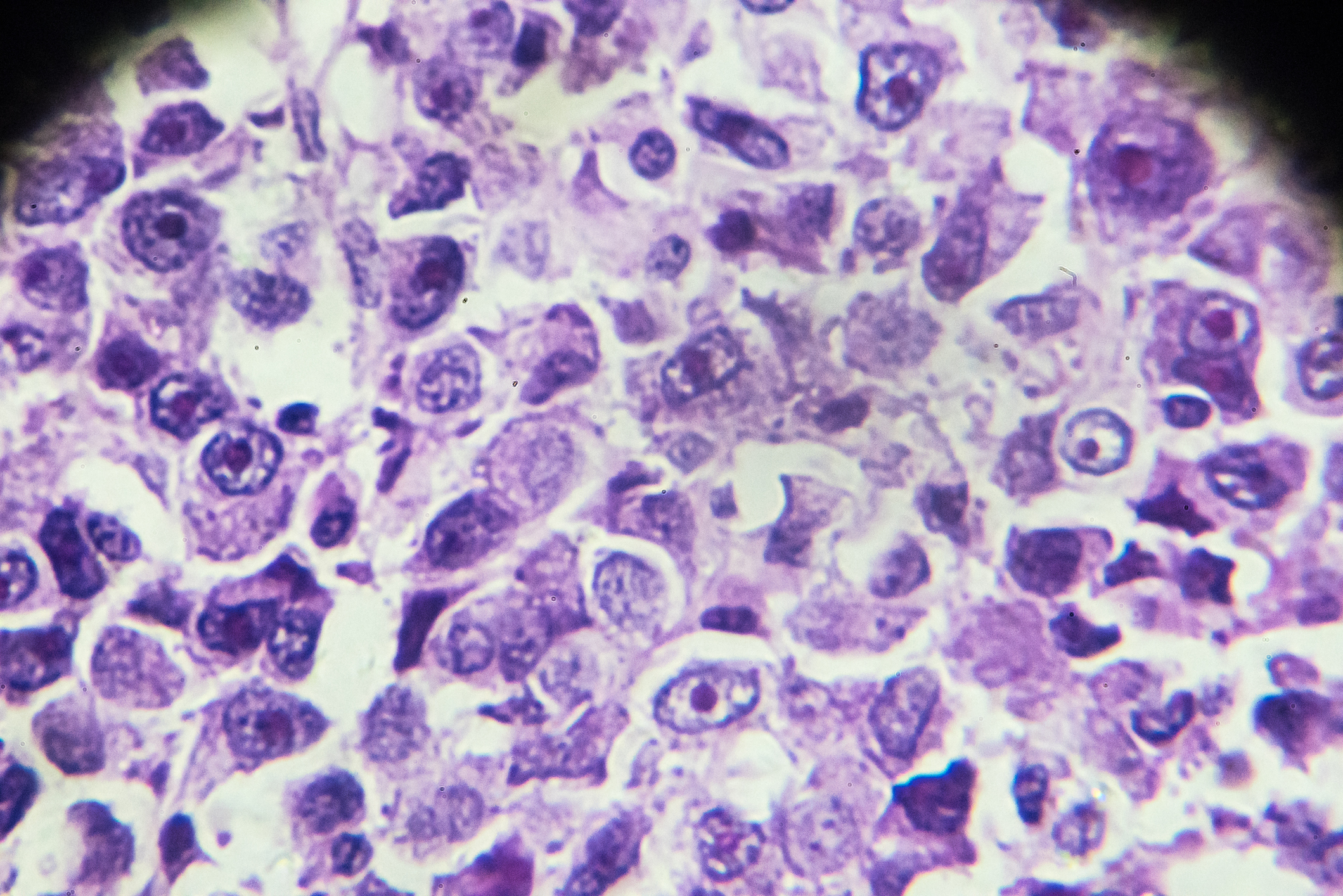

Abdominal pain is the most common initial symptom of pancreatic cancer, occurring in around half to two thirds of affected patients. Symptoms such as nausea and vomiting, lack of appetite, indigestion or weight loss only occur when the tumor hinders the production of digestive enzymes or their drainage into the duodenum or when it has already spread to other organs or the peritoneum.
Pancreatic carcinoma is a malignancy that is increasing in frequency. In 2021, there were 60,430 new cases in the USA, and the trend is rising. By 2030, carcinoma of the pancreas is expected to be the second most common malignant disease. In Europe, adenocarcinoma of the pancreas currently ranks tenth in carcinoma incidence [4], corresponding to 3% in each gender. Smoking poses the greatest risk of developing the disease, with dietary causes also contributing. An increase in prevalence is to be expected in the future due to the “fast food” generation [5,6]. The correlation between the intake of animal proteins and the occurrence of pancreatic cancer is significant.
At the time of initial diagnosis, 30-35% already have advanced local findings and 50-55% have metastasis. In the advanced stage, infiltration of the superior mesenteric artery and vein in particular limits surgical treatment. Only 10-15% of patients are still in a resectable stage of the tumor at the time of diagnosis. This is followed by adjuvant chemotherapy. The mean survival rate in these cases is 54.4 months [2,3,7]. This contrasts with a median survival rate of around 6 months in patients with inoperable local findings, vascular and plexus herniation and metastases.
In many cases, patients with pancreatic cancer experience considerable pain symptoms. Enzyme insufficiency, stenosis of the choledochal duct, infiltration of the adjacent parts of the intestine and the coeliac plexus are responsible for this [1]. The pain is often accompanied by persistent nausea, followed by weight loss. Pain management is one of the most important symptomatic measures in therapy.
X-ray examinations are of no value in the diagnosis of pancreatic carcinoma.
Sonography raises the suspicion of a tumor in any low-echo, irregularly limited pancreatic mass [9]. However, acute pancreatitis, which has a similar appearance on sonography, shows a different clinical and paraclinical situation with pain symptoms and laboratory findings.
Computed tomography (CT ) can show different image morphologies depending on the histological type, but it is not always possible to clearly differentiate between them. The criteria are listed in Table 1 . PET-CT has also established itself in the diagnosis of pancreatic cancer.

Magnetic resonance imaging also guarantees a high detection rate of pancreatic carcinomas. In addition, MRCP (magnetic resonance cholangiopancreatography) offers precise assessment of the efferent bile ducts and visualization of the Wirsungian duct. In recent years, the focus has also shifted to intraductal papillary mucinous neoplasia (IPMN), which can represent a precancerous condition [10].

Case study
Case study 1 (Fig. 1A to 1D) documents the course of palliative chemotherapy-treated pancreatic carcinoma in a 59-year-old patient. Initially, persistent nausea and slight weight loss were noticeable. Abdominal ultrasonography revealed an increase in tissue in the prevertebral lumbar region. The computer tomography requested for further diagnosis showed a locally invasive pancreatic head carcinoma and already extensive pulmonary metastasis. The considerable stenosis of the choledochal duct with blatant obstruction of the outflow was treated with a stent insertion. As the diagnosis was inoperable, she underwent several months of chemotherapy. The patient died 15 months after diagnosis.
Case 2 demonstrates (Fig. 2A to 2C) a carcinoma of the pancreatic head with a diameter of approx. 5 cm in a 70-year-old with pathological widening of the ductus Wirsungianus to 8 mm as well as overgrowth of the V. portae and peripancreatic fatty tissue infiltration. Liver filiae were already detectable.

Case 3 shows a carcinoma in the tail of the pancreas in a 70-year-old patient; the local calcifications indicate pre-existing chronic pancreatitis. Numerous liver metastases were already present.

Case 4 shows a pancreatic carcinoma in the corpus with walling of the mesenteric vascular root and infiltration of the adjacent parts of the small intestine. Small regional nodular structures were suspicious for local filiae of the lymph nodes.
Take-Home-Messages
- Pancreatic carcinomas are among the most aggressive malignancies. If symptoms occur, curative therapy is often no longer possible.
- Smoking and long-term malnutrition significantly increase the risk of the disease.
- In addition to laboratory chemical examinations, also under differential diagnostic aspects, imaging examination methods are essential for the diagnosis.
- Sonography, computed tomography and magnetic resonance imaging are used, PET-CT and MRCP are supplementary tools.
Literature:
- Coveler AL, et al: Pancreatic Cancer-Associated Pain Management. Oncologist 2021; 26(6): e971-972.
- Park W, Chawla A, O’Reilly EM: Pancreatic Cancer: A Review. JAMA 2021; 7; 326(9): 851-862.
- Smithy JW, O’Reilly EM: Pancreatic Cancer: Therapeutic trials in metastatic disease. J Surg Oncol 2021; 123(6): 1475-1488.
- De Baud F, Cascino S, Gatta G: Cancer of Pancreas. Crit Rev Oncol Hematol 2004; 50(2): 147-155.
- Nishi M: Pancreatic cancer. Gan To Kagaku Ryoho 2001; 28(2): 159-162.
- Benhamou S, Clavel F, Rezvani A, Doyon F: Relation between mortality in cancer of the pancreas and food and tobacco consumption in France. Biomed Pharmacother 1982; 36(8-9): 389-392.
- Gupta R, Amanam I, Chung V: Current and future therapies for advanced pancreatic cancer. J Surg Oncol 2017; 116(1): 25-34.
- Burgener FA, Herzog C, Meyers AP, Zaunbauer W: Differential diagnoses in computed tomography. 2nd, completely revised and expanded edition. Georg Thieme Verlag Stuttgart, New York: 1997; pp. 788.
- Seitz, Karlheinz et al: Klinische Sonographie und sonographische Differenzialdiagnose, 2008: DOI: 10.1055/b-0034-80131.
- Rummeny E: Pancreatic carcinoma – rare and insidious”, https://healthcare-in-europe.com,(last accessed 26.02.2024)
FAMILY PHYSICIAN PRACTICE 2024; 19(3): 46-48