Three countries, three conference presidents and three desired lectures on current research topics – that was on the agenda at the Presidential Symposium of the Epilepsy Conference in Interlaken. There was a state of the art on focus searching in epilepsies and information on the added value of MRI to malformations and genetics of cortical development. That zebrafish offers great prospects for pharmacological research was also reported.
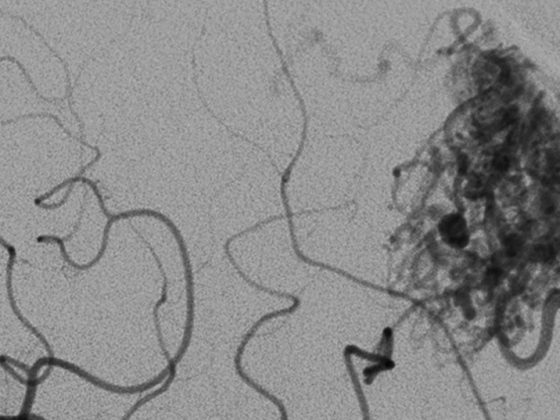
The conference president, Dr. med. Günther Krämer, Zurich, had invited Prof. Dr. med. Christoph M. Michel, Geneva, on the topic “Combination of electrical and hemodynamic imaging in epilepsy diagnostics” to the Epilepsy Conference in Interlaken. This made it clear that during an epileptic seizure there are maximum dynamics in the processes in the brain, but until now the methods were not sufficient to ensure temporal resolution. In contrast, new devices such as the 265-channel EEG or the 306-channel magnetoencephalogram (MEG) allow very high temporal resolution, and the combination of the two is now sufficiently fast. Together with mathematical models (Electric Source Imaging, ESI), reliable mapping and imaging can be performed today. In pre- and postoperative diagnostics, thanks to ESI, the initial site of seizures is correctly localized in 90% of cases. This method is advantageous due to good availability and for children.
Further improvements were obtained by combining EEG-fMRI and ESI-fMRI. Spike-map-informed-fMRI is an option in very difficult cases to reach an explanation for seizure phenomena after all. Prof. Michel also raised the issue that so far little is known about how a seizure stops. “The answer is more complicated than anyone thought. In the future, EEG and fMRI are appropriate for these types of research questions.”
Genetics of brain development
Giorgi Kuchukhidze, MD, is currently based in Innsbruck, Austria, and is an expert in the study of brain maturation and its disorders. He gave a presentation on MRI findings and underlying genetics. According to Dr. Kuchukhidze, about 90 genes have been described so far that, alone or in combination, may be responsible for CNS developmental disorders. For example, classic lissencephaly is associated with cerebellar atrophy, and two genetic forms have been identified in polymicrogyria: the bilateral peri-sylvian form with a gene locus in region Xq28 and the bilateral frontoparietal form with mutations in the GPR56 gene (G-rotin-coupled receptor 56) in region 16q13. In addition, one genetic defect can lead to different phenotypes, which complicates diagnosis. Genes for cytoskeleton formation are important for cell division, for example. If there are errors here, microcephaly may occur. The classification of developmental disorders is no longer usefully done at the gene level, but is instead arranged in functional groups such as tubulopathies or mitochondriopathies.
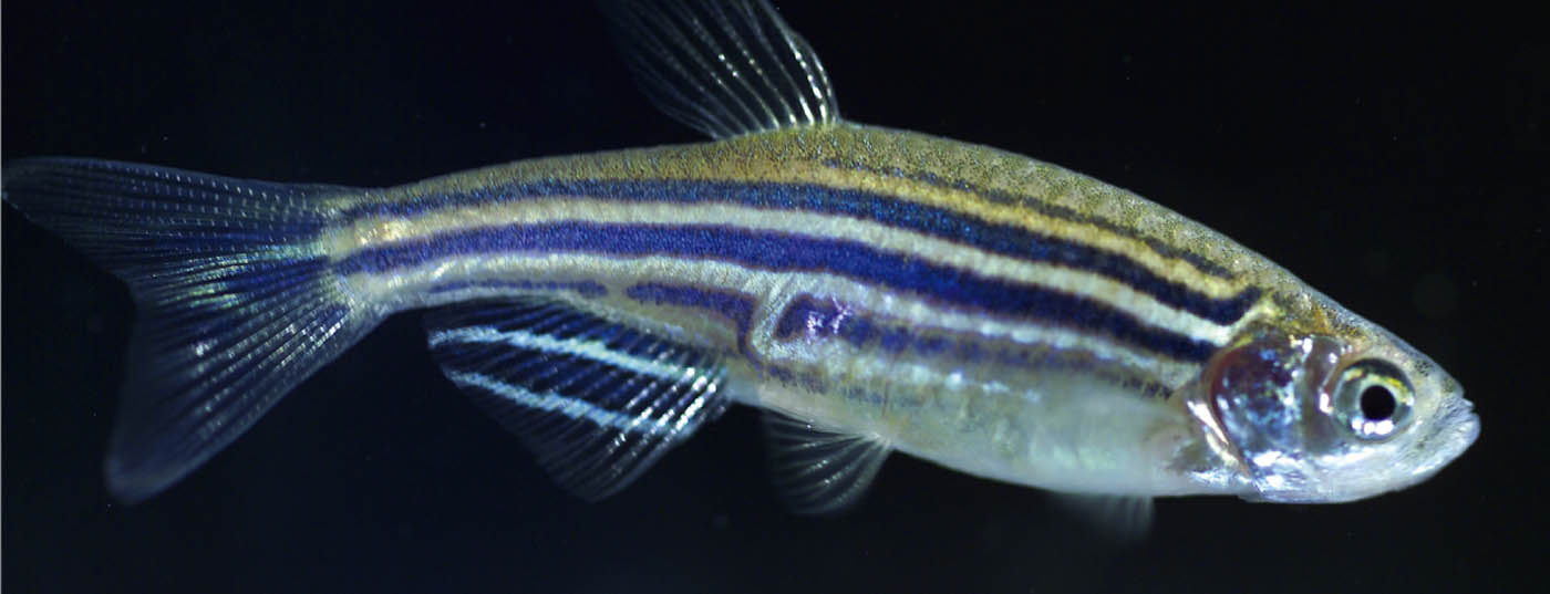
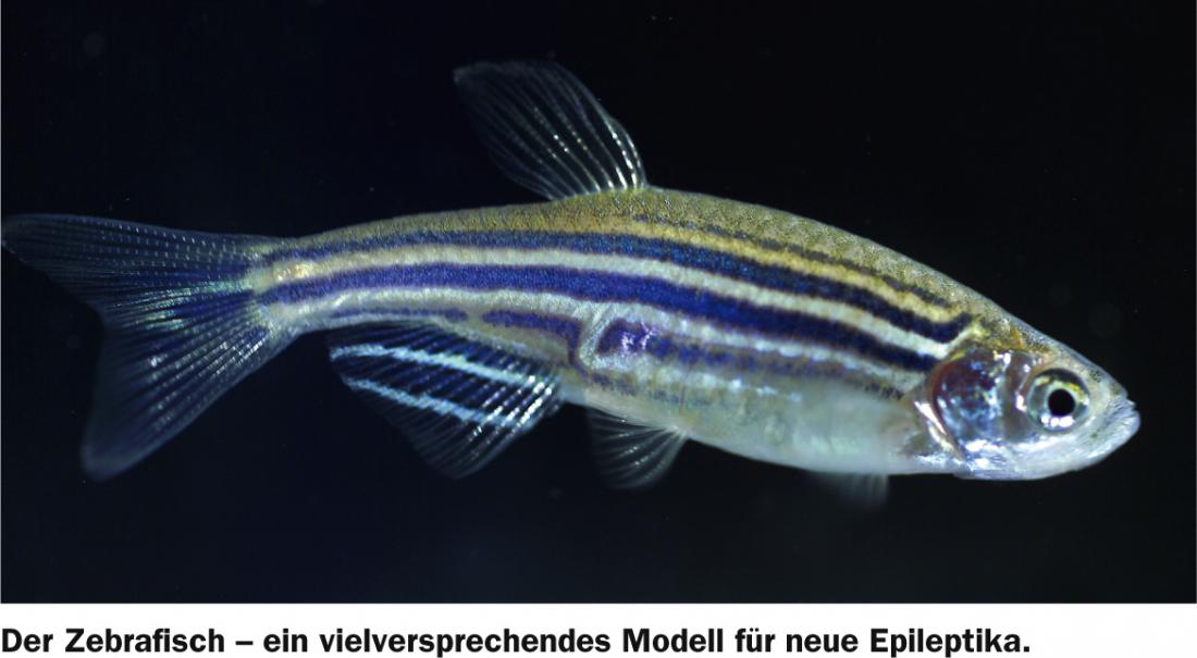
Learning from the zebrafish
Alex Crawford, MD, from Leuven, Belgium, spoke about “Genetic and chemoconvulsive zebrafish models for the development of new antiepileptic drugs.” The goal of Dr. Crawford’s work is to develop an animal model that can provide information about different seizures and the exact mechanisms involved, as well as demonstrate therapeutic success with antiepileptic drugs. Zebrafish have shown great promise in this regard, as the larvae already have a developed spinal column and a fully developed animal hatches within 24 hours. In addition, plant substances, such as those derived from turmeric, will be tested for their anticonvulsant efficacy. Dr. Crawford’s assessment of the future prospects of the research: “The prospects are great.”
Source: 8th Three-Country Meeting on Epilepsy, May 8-11, 2013, Interlaken.