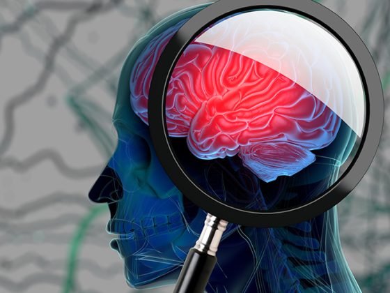
Using data from the longitudinal “UK Biobank” initiated in the pre-pandemic era, cerebral MRI findings before and after COVID-19 were collected for the first time in the same individuals and compared with a control group of uninfected individuals [1]. As a result, intermediate SARS-CoV-2 infected individuals showed a decrease in gray matter in the orbitofrontal cortex and a decrease in total brain mass. In those affected, cognitive test scores also deteriorated over time. Whether these changes are reversible is currently an open question. Another study [2] showed an increased rate of de novo dementia after COVID-19 compared with other pneumonias.
Many studies already showed COVID-19-associated abnormalities of brain structure. However, it has remained unclear whether milder courses of SARS-CoV-2 infection can also lead to such changes. A study has now been published in the prestigious journal Nature [1], which for the first time investigated cerebral MRI changes in SARS-CoV-2 infected individuals from whom cerebral MRI was available prior to the pandemic, as part of the large, longitudinal “UK Biobank Imaging Study” [2]. In the UK Biobank Imaging Study, which began in 2006, more than 40,000 people (>45 years) have since undergone multimodality cerebral MRI scanning at four centers using standardized protocols. The study was initially paused due to the pandemic; participants were then started to be invited for another MRI scan in February 2021. In the meantime, many of them had undergone SARS-CoV-2 infection.
To investigate the potential impact of SARS-CoV-2 infection on brain structure, the two scans (before and after COVID-19) were compared with participants not affected by COVID-19. The availability of preinfection imaging minimized the likelihood that unknown preexisting risk factors or abnormalities were later misinterpreted as COVID-related. Also, participants with incidental cerebral findings in the first scan were excluded from the study. The groups were comprehensively matched, ie, there were no significant differences in age, sex, ethnicity, mean blood pressure, diabetes mellitus, weight/BMI, alcohol and nicotine use, or socioeconomic status (“Townsend Deprivation Index”).
Of 785 eligible individuals in the biobank (ages 51-81) with two cerebral MRI scans each, 401 had experienced SARS-CoV-2 infection between the two scans, 15 of whom had been hospitalized. There was an average of 141 days between the diagnosis of infection and the second scan. The control group included 384 individuals. The interval between the two brain scans averaged 3.2 ± 1.6 years in both groups.
As a result, there were significant longitudinal effects or MRI changes in the group of intermediate SARS-CoV-2 infected individuals. These included a decrease in gray matter and a decrease in tissue contrast in the orbitofrontal cortex (cortex in the frontal area over the orbits) and in the so-called parahippocampal gyrus (part of the limbic system located in the temporal lobe). There were also tissue changes or damage in brain regions functionally associated with the primary olfactory cortex and a greater decrease in total brain mass. Those infected with SARS-CoV-2 in the interim also showed significantly more deterioration (in the time between the two scans) in cognitive tests than noninfected individuals. These longitudinal group differences (in imaging and cognition) persisted even when the 15 participants hospitalized for COVID-19 were not included in the statistics.
The pathomechanism of SARS-CoV-2-associated brain changes now requires further research. The researchers discuss a spread of the virus via olfactory-neuronal pathways and inflammatory processes. The loss of sensory-olfactory input due to the loss of the sense of smell (anosmia) may also have indirectly caused structural changes, the authors of the study said.
“The UK Biobank data show that there is a morphological correlate for post-COVID neurological symptoms,” commented Prof. Peter Berlit, MD, Secretary General of the DGN. “Whether the changes documented in the imaging are reversible during the course or persist in the sense of neurodegeneration in the long term must now be further investigated in the follow-up.”
Another study [3] also described COVID-19-associated functional cerebral changes. Here, however, the more than 10,000 people affected all had SARS-CoV-2 pneumonia with a severe course. New-onset dementia developed in 3% after >30 days. The risk of dementia after SARS-CoV-2 pneumonia was 30% higher (OR 1.3) than non-COVID-19-associated pneumonia in this study. New-onset dementia was defined using primary diagnosis codes according to ICD-10-CM (F01.5, F02.8, F03.9, G30, G31, G32). Affected individuals with documented preexisting dementia symptoms or cognitive deficits were excluded. Comorbidities that may increase the risk of developing dementia were considered in the multivariate analysis (e.g., hypertension; drug, nicotine, and alcohol use; certain neurologic and psychiatric disorders).
“The data show that the virus, although fortunately only in rare cases, can also lead to changes in the brain in the long-term course. Against this background, vaccination offers protection not only against severe acute courses of the infection, but also against secondary damage,” concludes the expert.
Literature:
- Douaud G, Lee S, Alfaro-Almagro F et al. SARS-CoV-2 is associated with changes in brain structure in UK Biobank. Nature 2022 Mar 7. doi: 10.1038/s41586-022-04569-5. online ahead of print.
- https://www.ukbiobank.ac.uk/explore-your-participation/contribute-further/imagin…
- Qureshi AI, Baskett WI, Huang W et al. New Onset Dementia Among Survivors of Pneumonia Associated with Severe Acute Respiratory Syndrome Coronavirus 2 Infection. 2022 Infectious Diseases Society of America. https://watermark.silverchair.com/ofac115.pdf
Original publication:
doi: 10.1038/s41586-022-04569-5