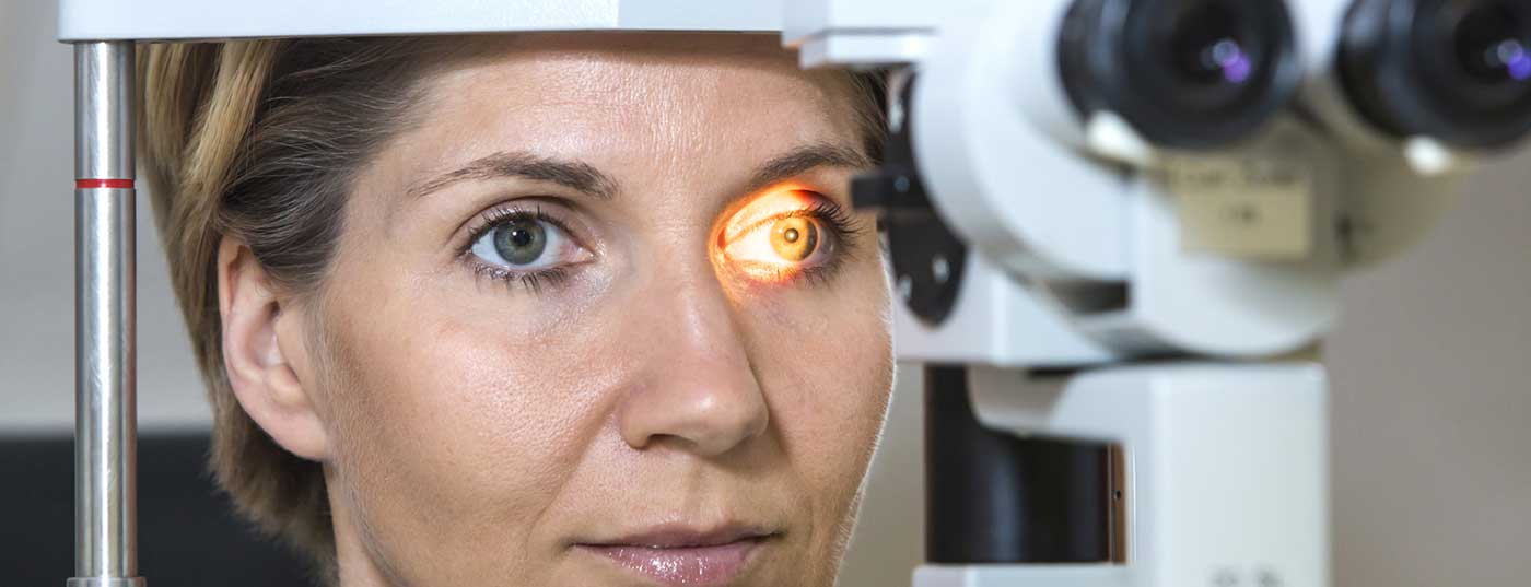
Funduscopy and further diagnostic procedures can detect diabetic retinopathy at an early stage. Today, innovative treatment options exist that can slow further progression. Diabetics should be examined ophthalmologically at regular intervals. This is because a deterioration in vision only becomes noticeable to many sufferers at an advanced stage.
Diabetic retinopathy is a common, progressive microvascular complication of diabetes mellitus. Depending on the study, one in four to one in five diabetics develops diabetic retinopathy. Risk factors include duration of diabetes, glycemic control (HbA1c), and arterial hypertension. Vision loss in diabetic retinal complications usually develops gradually and, with the exception of vitreous hemorrhage, is not associated with pain. If left untreated, diabetic retinopathy can lead to blindness. “Screening is very very important,” emphasized Felix Rombold, MD, ophthalmologist, Augsburg Eye Hospital [1].
Fluorescence angiography and optical coherence tomography (OCT)
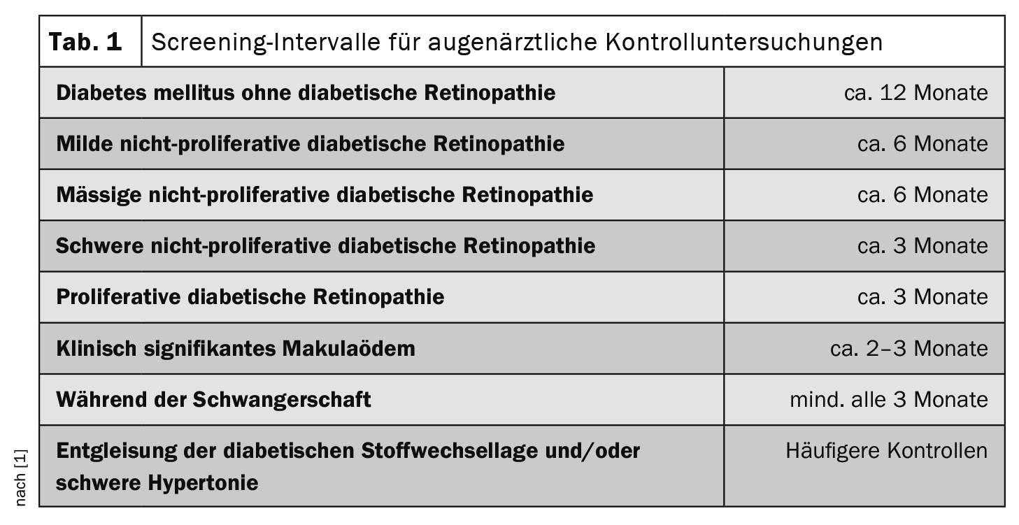
Regular eye examinations (Table 1) can detect morphologic changes even before functional impairment occurs, so that therapeutic measures can be initiated in time. While type 1 diabetics often present with ischemia but no visual loss, type 2 diabetics focus on macular edema. Clinical manifestations are distinguished as non-proliferative (NPDR) from proliferative diabetic retinopathy (PDR). The extent of retinal involvement can be assessed using the Diabetic Retinopathy Severity Scale (DRSS) and the Early Treatment Diabetic Retinopathy Study-DRSS (ETDRS-DRSS) [2,3]. “If peripheral ischemia or neovascularization is suspected, we do advanced diagnostics with fluorescein angiography,” Dr. Rombold explained [1]. Fluorescence angiography can be used to detect diabetic vascular changes such as microaneurysms and microperfusion defects as well as vascular leakage. Accurate analysis of retinal edema is made possible by optical coherence tomography (OCT). Especially in regions with a low density of ophthalmologists, the importance of artificial intelligence applications is also increasing. Using fundus photographs, anatomically relevant structures such as the macula, optic nerve or blood vessels are identified by an algorithm and the entire image acquisition is examined for pathological diabetes-related changes, such as microaneurysms.
Intravitreal surgical drug application
To preserve vision, the best possible blood glucose control and treatment of the underlying disease is essential. Of the targeted therapies available today, intravitreal surgical medication (IVOM) currently achieves better visual outcomes than laser therapy [1].
Diabetic macular edema: anti-VEGF therapy
Currently approved anti-VEGF antibodies in Switzerland for diabetic macular edema, which are injected into the vitreous by IVOM, include aflibercept (Eylea®), and ranibizumab (Lucentis®) [4]. Bevacizumab (Avastin®) can only be used “off-label” [4]. With these drugs, a great increase in visual acuity can be achieved, the speaker reported. In the first half of the year, the active ingredients must be administered every four weeks, which requires a certain degree of patient adherence. If persistent vitreous hemorrhage or traction amotio occurs, vitrectomy is indicated. This involves removing the vitreous, detaching the existing traction membranes from the retina, and treating the retina with endolaser coagulation to prevent further bleeding and proliferation. Subsequently, the glass body is replaced by an air-gas mixture or silicone oil.
Congress: Diabetology without borders
Literature:
- “Diabetic Retinopathy: Retinopathy Screening Using Artificial Intelligence & Ophthalmic Quality Control,” Felix Rombold, MD, Diabetology Limitless, Feb. 03, 2022.
- Davis MD, et al: Risk factors for high risk proliferative diabetic retinopathy and severe visual loss: early treatment diabetic retinopathy study report #18. Invest Ophthal mol Vis Sci 1998; 39(2): 233-252.
- Early Treatment Diabetic Retinopathy Study Research Group. Grading diabetic retinopathy from stereoscopic color fundus photographs – an extension of the modified Airlie House classi fication. ETDRS report number 10. ophthalmology. 1991; 98(5 suppl):786-806. doi:10.1016/S0161-6420(13)38012-9
- Drug Information, www.swissmedicinfo.ch,(last accessed Feb. 07 , 2023).
HAUSARZT PRAXIS 2023; 18(2): 34 (published 2/22/2013, ahead of print).


