Transthyretin amyloidosis with cardiomyopathy is a significantly underdiagnosed disease that often presents with clinical symptoms of heart failure. Due to the age pyramid, the number of patients will most likely increase. But a broad spectrum of symptoms makes diagnosis difficult. In this context, early diagnosis would be important in order to rapidly implement the existing treatment option.
Amyloidosis is a group of diseases characterized by deposition of pathological proteins in the interstitium. If the amyloid arises from deposition of misfolded transthyretin (TTR) monomers primarily in the heart and peripheral or autonomic nerves, it is referred to as transthyretin amyloidosis with cardiomyopathy (ATTR-CM). It has a high morbidity and mortality. The clinical picture is so multifaceted that ATTR-CM is often only discovered in advanced stages. A close exchange between primary care physicians, cardiologists, and nuclear medicine physicians is therefore desirable.
Typical characteristics
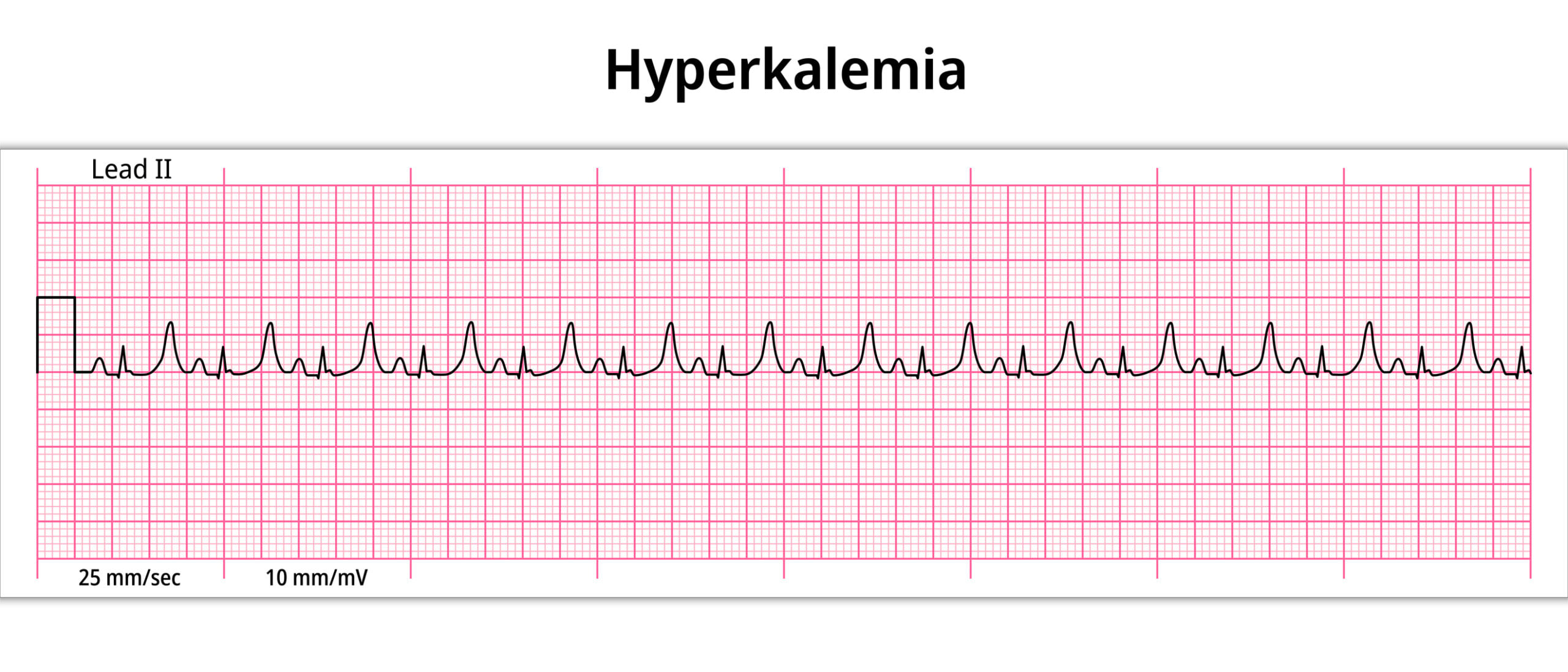
For example, in elderly male patients older than 60 years with heart failure with preserved ejection fraction (HFpEF), ATTR-CM should be considered if they do not respond to standard therapy with beta-blockers or ACE inhibitors. Bilateral carpal tunnel syndrome, elevated NT-proBNP levels, or cardiac wall thickening greater than 12 mm are also indicative of this form of amyloidosis (Table 1). A skeletal scintigraphy can bring clarity. Characteristic above all is a “black heart”.

Wild type or hereditary?
Destabilization of the TTR protrein and formation of misfolded TTR proteins result in the formation of amyloid fibrils, which can be deposited in various parts of the body, where they can lead to severe structural changes. In ATTR amyloidosis, peripheral nerves or the heart are mainly affected. Accordingly, a distinction is made in ATTR amyloidosis with polyneuropathy or ATTR-CM. The cause may be either a mutation in the transthyretin gene as a hereditary form or an age-related wild type. ATTR-CM usually occurs as a wild type, but can also manifest as mutation-related. It develops insidiously, but should be diagnosed early. This is because the untreated median survival from diagnosis is 3.6 years.
Diagnostics are performed on the basis of different measures. Echocardiography can detect thickened heart walls. In addition, pump function is impaired and, in later stages, the atria are dilated. A distinctive feature is the almost preserved or only slightly reduced longitudinal deformation in the apical segments of the left ventricle. Skeletal scintigraphy is also recommended for noninvasive screening.
Starting with the causes
Tafamidis, an oral inhibitor of TTR tetramer dissociation, is the first and so far only causal therapy available. It binds to the thyroxine binding site of the TTR tetramer, stabilizing it and preventing fibril formation. In the pivotal study, 30% lower all-cause mortality and 32% fewer cardiovascular-related hospitalizations were seen for the 20 mg and 4× 20 mg tafamidis meglumine dose arms compared with placebo.
The results of the open-label extension study now showed a significant survival benefit in patients receiving 80 mg of tafamidis meglumine versus those receiving 20 mg with a relative reduction in the risk of death of 30%. When corrected for relevant covariates, there was an overall relative reduction in mortality risk of 43%. The drug was well tolerated at both doses and the safety profile was comparable to placebo.
Further reading:
- Maurer MS, et al: Circulation 2017; 135:1357-1377.
- González-López, et al: Eur Heart J. 2015; 36(38): 2585-2594.
- Ruberg FL, et al: J Am Coll Cardiol. 2019; 73(22): 2872-2892.
- Maurer MS, et al: Circ Heart Fail. 2019; 12: e006075
- Donnelly J, Hanna M. Cleve Clin J Med 2017; 84(12 Suppl 3): 12-26.
- Aus dem Siepen, et al: Clin Res Cardiol. 2019; 108(12): 1324-1330.
- Gillmore JD, et al: Circulation 2016; 133: 2404-2412.
- Damy T, et al: Eur J Heart Fail 2020 Oct 18. doi.org/10.1002/ejhf.202.
- Yilmaz A, et al: Clinical Research in Cardiology, publ. online January 2021, https://doi.org/10.1007/s00392-020-01799-3
CARDIOVASC 2021; 20(2): 18