Research into the skin microbiome has become increasingly important in recent years. A study presented at the 2019 ADF Annual Meeting examined the effects of topical cortisone externals on the composition of the skin microbiome of atopic patients. In the process, interesting findings were discovered.
In skin diseases such as atopic dermatitis, a restricted diversity of the skin microbiome is measurable compared to healthy skin [1,2,5,6]. In acute phases of atopic eczema, increased colonization with Staphylococcus aureus (S. aureus) and displacement of other bacterial species occurs on affected skin areas. Microbial dysbiosis of inflamed atopic skin lesions has been studied several times. However, the body of evidence on host-microbiome interactions has been limited to date.
Colonization of S. aureus varies
In the present study, lesional and non-lesional skin samples of the elbows of atopic patients (n=7) were analyzed by molecular biology before and after treatment with a topical cortisone preparation (Dermatop®) [7] and compared with biopsies of a skin-healthy control group (n=8) [1,2] (Fig. 1). 16S amplicon sequencing was used with an Illumina MiSeq TM instrument [3], as well as “Rhea”, a set of R-scripts for microbial profile analysis [4].
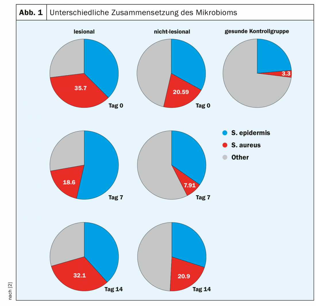
At baseline, the skin microbiome of biopsies in the lesional state contained a greater proportion of S. aureus than in the nonlesional state (35.7% vs. 20.59%). S. aureus colonization innon-atopic skin samples was significantly lower in comparison (<5%). Also distinct are the differences in commensal diversity, which is significantly higher in non-atopic biopsies. After 1 week of treatment with a Dermatop® [7], a decrease in S. aureus colonization was measurable (to 18.60% and 7.91%, respectively) in both the lesional and non-lesional skin microbiome. After 2 weeks of treatment, the percentage of S. aureus increased significantly in both lesional and nonlesional microbiome samples, to values similar to those at baseline (32.10% and 20.90%, respectively).
There is much need for research
How different therapeutic agents affect the skin microbiome in atopic dermatitis is among the many questions still unresolved. For example, it is still not known whether microbial changes in atopic dermatitis (e.g., increased S. aureus colonization) are a cause or consequence of epidermal barrier dysfunction and inflammatory processes [6]. As future treatment strategies, in addition to further development of current therapies, new approaches such as manipulation of the microbiome by topically applied commensals are discussed [5]. In any case, it remains exciting.
Source: ADF, Munich-Garching (D)
Literature:
- Amar Y, et al.: Skin microbiome diversity and transcriptome profiling in patients with atopic dermatitis. 46th Annual Meeting of the Arbeitsgemeinschaft Dermatologische Forschung (ADF). P015. Experimental Dermatology 2019; 28(3): e1-e122 https://onlinelibrary.wiley.com/doi/10.1111/exd.13859
- Amar Y, et al: Exploration of Microbiome Shifts in Dermatop treated atopic dermatitis patients. Poster presentation, ADF Annual Meeting, Munich-Garching, March 16 2019.
- MiSeqTM System, www.illumina.com/content/dam/illumina-marketing/documents/products/datasheets/miseq-data-sheet-770-2011-001-translations/
- miseq-specification-sheet-770-2011-001-deu.pdf
- Lagkouvardos I, et al: Rhea: a transparent and modular R pipeline for microbial profiling based on 16S rRNA gene amplicons. Peer J 2017; 5:e2836.
- Paller, et al: The microbiome in patients with atopic dermatitis. Journal of Allergy and Clinical Immunology 2019; 143 (1); 26-35.
- Kim JE, Kim HS: Microbiome of the Skin and Gut in Atopic Dermatitis (AD): Understanding the Pathophysiology and Finding Novel Management Strategies. Clin Med 2019; 8(4). pii: E444. doi: 10.3390/jcm8040444. Dermatop: Technical information. https://info.diagnosia.com/de-de/p/377917/dermatop
DERMATOLOGIE PRAXIS 2019; 29(4): 22