The International Parkinson and Movement Disorder Society (MDS) gathers thousands of clinicians, researchers, trainees and industry advocates with an interest in current research and approaches to the diagnosis and treatment of movement disorders at its Congress of Parkinson’s Disease and Movement Disorders® each year. The purpose is to share ideas, generate interest among all stakeholders, and advance clinical and scientific disciplines.
Narcolepsy with cataplexy, also known as narcolepsy type 1 or NT1, is a sleep disorder characterized by excessive daytime sleepiness, hypnagogic hallucinations, sleep paralysis, and cataplexy (loss of muscle tone triggered by strong emotions). It is caused by a deficiency of the hypothalamic neuropeptide hypocretin/orexin. The underlying mechanism of the disease remains unknown. However, currently available data support an immunologic mechanism leading to loss of hypocretin-producing neurons. In addition, environmental risk factors and genetic variations at multiple loci have been shown to be associated with NT1. The HCRT gene encodes hypocretin/orexin and is the only phenotype documented under this gene in OMIM to date. To gain further insight into HCRT-related narcolepsy, results were presented from five additional patients from two unrelated families who have a homozygous truncated variant in this gene [1]. Whole-exome sequencing identified a common homozygous variant of HCRT in all five patients. Sanger sequencing confirmed the presence of this variant as homozygous in all affected individuals and as heterozygous or absent in the available unaffected family members who underwent the test.
Clinically, the patients had the features of cataplexy: Episodes characterized by seizures of axial atonia with preserved consciousness. These seizures were typically triggered by strong emotions. Seizures began at 3-4 months of age, lasted between 10 and 50 seconds, and occurred up to 12 times per day. Neurologic examination performed between seizures revealed unremarkable findings. The onset of excessive daytime sleepiness occurred at a mean age of six months, with seizures lasting up to 40 minutes and recurring two to three times daily. At baseline, two patients exhibited mild motor delay. EEG and MRI results of the brain were normal. Measurement of hypocretin-1 level in CSF by direct radioimmunoassay showed a significant reduction with values below 90 pg/ml (normal >200 pg/ml). As for follow-up, the parents refused any treatment. Over time, cataplexy improved markedly in all patients: seizures disappeared completely in two patients by two years of age, in one patient by four years of age, and significant improvement was seen in the remaining two patients. Regarding narcoleptic episodes, two patients recovered completely, one patient showed marked improvement, and two patients showed mild improvement. Development was normal in all patients except one, who showed a mild motor delay.
Narcolepsy type 1 is characterized by a deficiency of the hypothalamic neuropeptide HCRT. However, the exact pathological mechanism responsible for the loss of hypocretin neurons in most narcolepsy patients with cataplexy remains largely unknown. However, the strong association between the HLADQB1*06:02 allele, polymorphisms in other immune-related genes, and the immunologic-autoimmune response observed in narcolepsy patients with cataplexy supports the autoimmune hypothesis. Therefore, genetic testing of the HCRT gene should be performed in certain subgroups of NT1, particularly those with early onset, familial cases, and a predominantly cataplexic phenotype.
Olfactory function as a biomarker for Parkinson’s disease
Hyposmia is a common symptom in the early stages of PD. Therefore, the aim of one study was to improve the discrimination between patients with Parkinson’s disease (PD) and non-PD controls by combining the real-time quaking-induced conversion (RT-QuIC) test using olfactory mucosa (OM) with clinical data on olfactory function [2]. For this purpose, samples of olfactory epithelium were collected from subjects with early Parkinson’s disease (PD) (n=10) and from control subjects not suffering from PD (n=10). To ensure applicability in real-world clinical settings, controls were defined as individuals with parkinsonism whose nigrostriatal dopaminergic nerve terminals were preserved according to PET imaging, suggesting that symptoms were caused by other factors or a precursor stage of nigrostriatal degeneration. The ability to induce a-synuclein aggregation of OM samples containing human recombinant alpha-synuclein was investigated using the RT-QuIC assay.
Olfactory function test did not differ between PD patients with negative and positive RT-QuIC results (total score 20.3 versus 19.1). This trend was also observed in non-PD control subjects according to RT-QuIC results (18.5 versus 19.6). The combination of olfactory function and RT-QuIC assay using olfactory epithelium did not provide satisfactory results. However, it should be noted that the non-PD control subjects were not healthy; they were significantly older than the PD patients and may have had subclinical degenerative disorders that could have influenced RT-QuIC results. Moreover, the PD patients were at an early stage of the disease, so longitudinal follow-up could clarify their diagnosis.
The role of niacin
Daily intake of niacin in Parkinson’s disease patients may lead to a reduction in UPDRS-III scores. Niacin can act in a number of ways in a cell. It is anti-inflammatory when it acts through its receptor GPR109A or HCAR-2. Niacin increases NAD levels, promoting mitochondrial functions. It was also shown that phosphorylation of α-synuclein at serine residue 129 decreased in Parkinson’s disease patients taking 250 mg of niacin daily for six months, whereas these levels were increased in exosomes in the niacin group compared with the placebo group. Now to investigate whether exosomal α-synuclein in plasma is an indicator of prognosis in PD and whether low-dose daily niacin administration might be helpful.
The randomized trial of daily 250 mg niacin or placebo lasted six months, with both groups taking free-release niacin for the subsequent six months [3]. Total plasma exosome levels did not differ significantly between groups or time points. However, oligomers decreased by 1.7 ng/ml from baseline (BL) to month 6 in the placebo group. Significant changes were observed in the placebo group for the ratio of exosomes to plasma p(s129) α-syn and oligomers. The ratio of oligomers to exosomes in plasma increased during open niacin treatment. However, in the placebo group, oligomers decreased by 1.6 ng/ml. In addition, the concentration of L1 cell adhesion molecule (L1CAM) in plasma exosomes decreased by 0.14 ng/ml in the placebo group. In conclusion, lower levels of aggregate-forming α-synuclein and modulated exosome-associated α-synuclein in the periphery after niacin supplementation in PD suggest disease modification that may underlie the symptom improvements observed in previous studies.
Non-motor symptoms in dystonia
GTP-cyclohydrolase 1-deficient dopa-responsive dystonia (GTPCH1-deficient DRD) is a rare disorder characterized by lower-limb dystonia occurring at a young age with a dramatic and sustained response to low-dose oral levodopa. There is limited and conflicting evidence based on small case series describing the frequency and severity of nonmotor symptoms. Therefore, patients with confirmed GTPCH1-deficient DRD were prospectively recruited from a tertiary movement disorder clinic or a North Bristol NHS Trust research database. Subjects were assessed for a range of non-motor symptoms using validated questionnaires, with a focus on mood disorders, sleep and cognition [4].
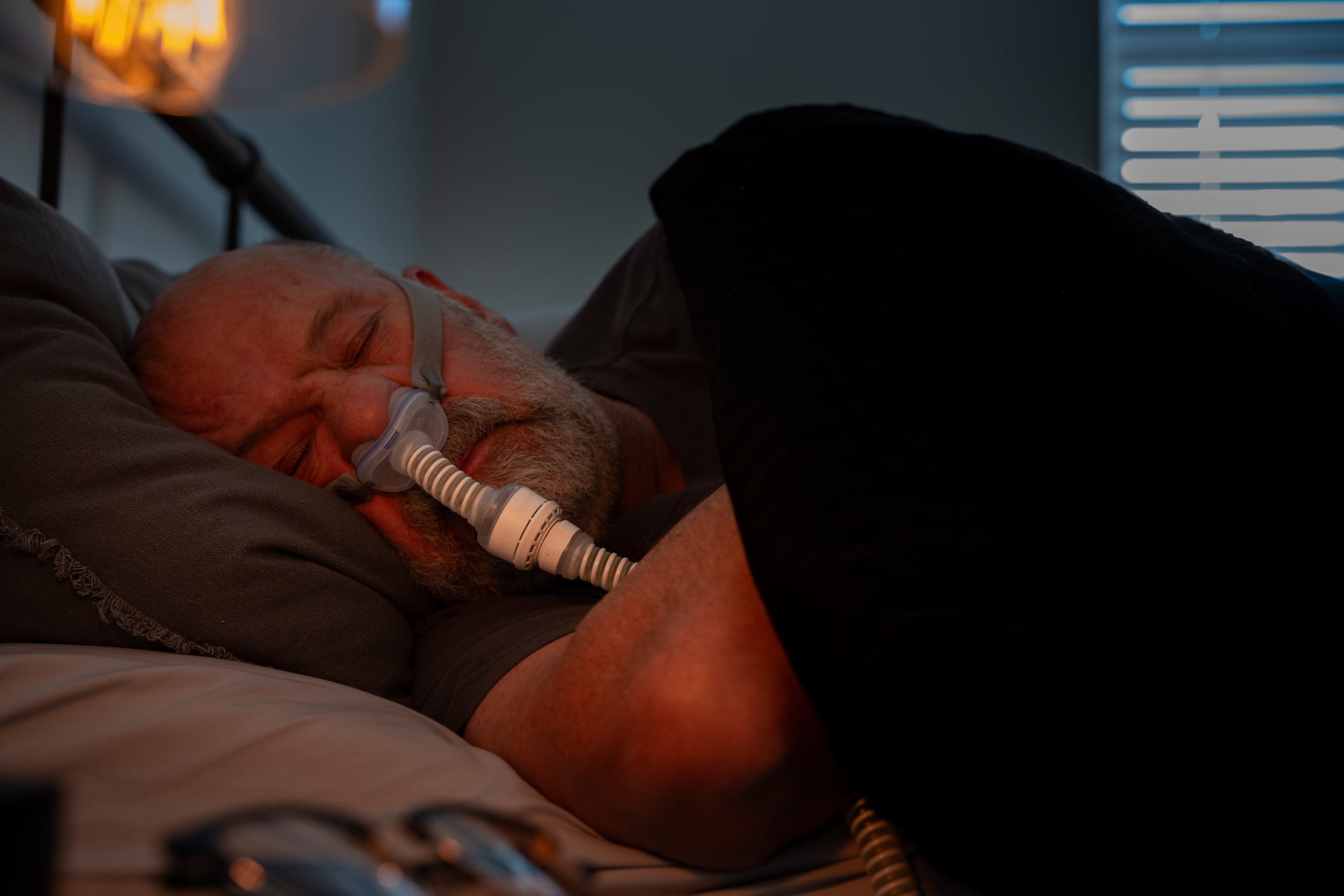
11 patients (female = 7; male = 4) aged 26 to 75 years were recruited from six families. The median age at diagnosis was 17 years. 91% had a dystonic phenotype at presentation and 1/11 had adult-onset parkinsonism. 10/11 (91%) of patients were taking levodopa at a mean levodopa equivalent dose of 222 mg/day. Motor symptoms were well controlled in all patients. 6/11 (55%) were taking an antidepressant. Comorbid depression was identified in 73% of participants, anxiety in 18%, obsessive-compulsive disorder (18%), autism spectrum disorder in 2/11 (18%), and mild learning disabilities in 1/11. 18% had a diagnosis of obstructive sleep apnea.
In this cohort of patients with GTPCH1-DRD deficiency, the burden of non-motor symptoms is high, particularly depression and sleep disturbances. In addition, there may be unrecognized mild cognitive dysfunction. More in-depth phenotyping with neuropsychometric and objective sleep measures is warranted, as these additional symptoms may affect quality of life and be amenable to treatment.
Congress: International Congress on Parkinson’s Disease and Movement Disorders 2023
Literature:
- Hakami W, Alshahwan S, Alkuraya F, Tabarki B: Bi-allelic variants in HCRT cause autosomal recessive narcolepsy. LBA-1st International Congress of Parkinson’s Disease and Movement Disorders® 2023.
- Pham N, Kwak I-H, Ma H-I, Kim Y: Olfactory mucosa RT-QuIC in combination with olfactory function as a biomarker for Parkinson’s disease. LBA-2nd International Congress of Parkinson’s Disease and Movement Disorders® 2023.
- Wakae C, Seamon M, Hannah Y, et al: Is plasma exosomal α-synuclein prognostic in Parkinson’s disease (PD) patients? Role of niacin supplements. LBA-4th International Congress of Parkinson’s Disease and Movement Disorders® 2023.
- Morrison H, Blackman J, Gabb V, et al: Non-motor symptoms in patients with GTPCH1-deficient dopa-responsive dystonia. LBA-17th International Congress of Parkinson’s Disease and Movement Disorders® 2023.
InFo NEUROLOGY & PSYCHIATRY 2023; 21(5): 22-23. (published 10/22-23, ahead of print)