New developments in computer-aided data classification, machine learning methods, development of specific tracer substances, and clinical application of high-field MR technology at field strengths <3 Tesla are leading to increasingly rapid developments in epilepsy imaging – an overview.
Imaging evaluation of patients after a first-time seizure serves
- evidence of a treatable cause in a provoked seizure in the context of another underlying disease or cause of seizure,
- the detection of a structural epileptogenic lesion for prognostic assessment,
- The planning of epilepsy surgery for pharmaco-resistant epilepsy.
The method of choice is MR examination at a field strength of 3 Tesla with a dedicated epilepsy protocol. In clinically and neurophysiogically clear idiopathic epilepsy syndromes (e.g., absence epilepsy), imaging may not be necessary.
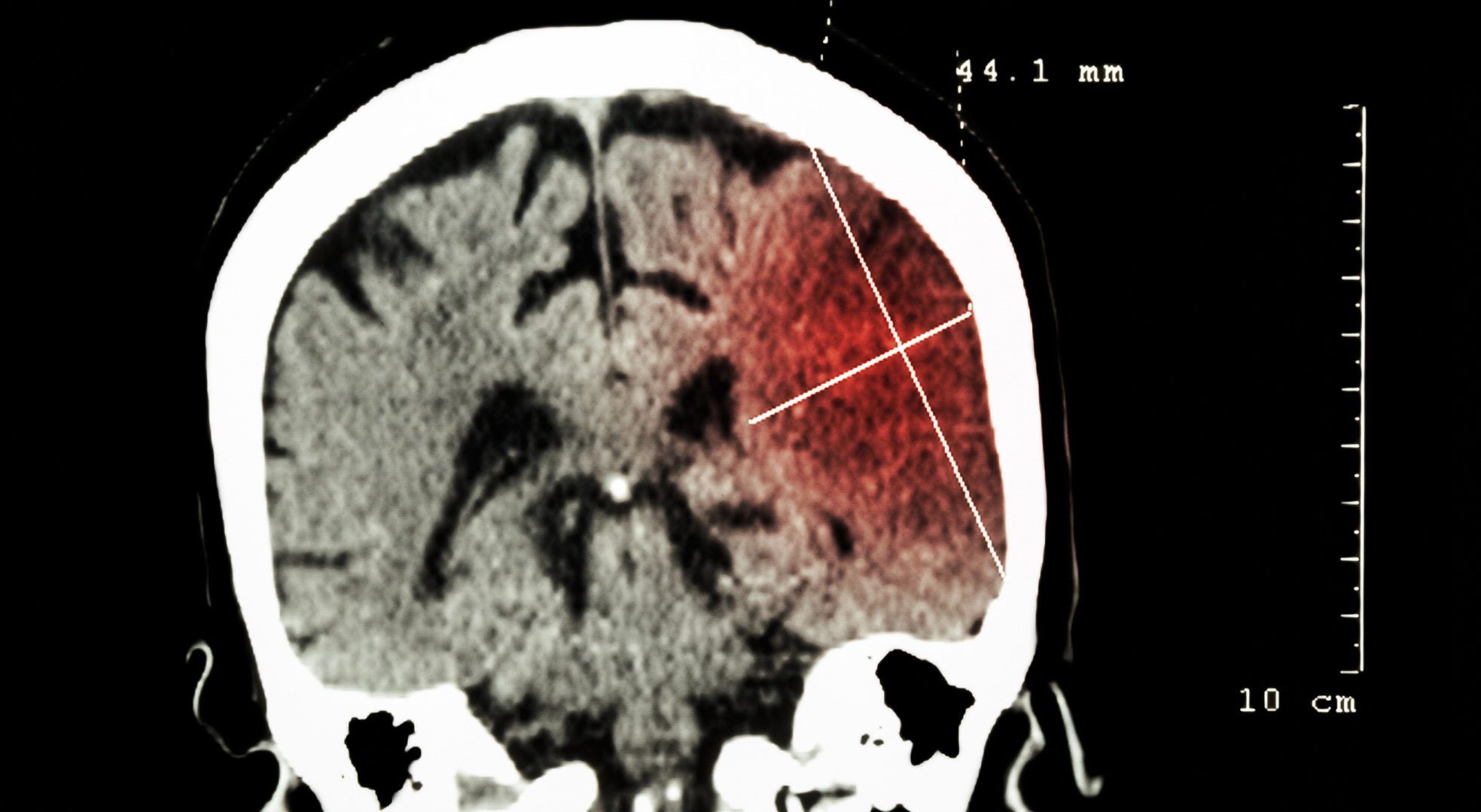
Postprocessing and reformatting techniques can aid visual image analysis, especially for subtle findings (epileptogenic malformations of cortical development). Functional imaging techniques (PET, fMRI, brain perfusion measurements with SPECT or ASL) have an additive value to clinical seizure analysis and neurophysiology for localization diagnosis and preoperative planning under the principle of “converging evidence” (Fig. 1).

The detection of a structural epileptogenic lesion has different significance at different stages of the diagnostic workup of epilepsy (Overview1). A “structural epileptogenic lesion” is defined as an alteration of the brain detectable macroscopically by CT or MRI that has an epileptogenic effect “per se” (such as a malformation of cortical development) or may have a secondary epileptogenic effect by causing hyperexcitability of the surrounding brain area (such as a hemorrhagic cavernoma). Especially after the occurrence of a first epileptic seizure, changes in the brain that may be the cause of a symptomatic epileptic seizure (such as a meningioma or sinus vein thrombosis) must therefore be distinguished from changes that may be the causal cause of an onset of epilepsy (such as a permanent glian scar after a stroke, traumatic brain injury, or previous infection of the brain). Imaging localization diagnosis after a first seizure therefore follows the principle “risk does not equate causation”, i.e. not every structural change of the brain is necessarily epileptogenic (e.g. venous malformations, arachnoid cysts). Conversely, in patients who develop epilepsy after an epileptic seizure, depending on the underlying epilepsy syndrome, structural changes are also recorded in visual analysis in about 11-28% of cases – in focal epilepsy, the percentage is much higher, exceeding 50% [1,2]. In epilepsy in which seizures cannot be suppressed despite drug treatment (approximately one-third of cases), it should be determined whether there is a structural epileptogenic lesion requiring epilepsy surgery.

MR Imaging
Diagnostic imaging techniques for the detection of structural epileptogenic lesions and for functional localization diagnosis have steadily gained importance in the last decades, especially in pre-surgical epilepsy diagnostics [3]. Technological advances in image acquisition, the widespread provision of medium field strength magnetic resonance imaging (3 Tesla) and, more recently, the clinical approval of ultra-high field magnetic resonance imaging (7 Tesla) have led to an ever-increasing increase in the information content of image datasets. Contributing factors include developments in new MR hardware (multichannel and surface head coils), new MRI sequences (Table 1) such as susceptibility-weighted imaging (SWI), and new experimental MR technologies (simultaneous EEG-fMRI) [4,5]. These new technologies, in combination with optimized epilepsy protocols according to the recommendations of the International League against Epilepsy (ILAE) [6–8] allow improved detection of structural epileptogenic lesions in refractory epilepsies in up to 80% of cases [9,10]. SWI, a flow-compensated, spatially high-resolution 3D gradient echo sequence technique, allows visualization of microhemorrhages, cavernomas, and calcifications of the cortex. Diffusion-weighted and perfusion-assisted imaging techniques (KM-assisted perfusion imaging and noncontrast measurements of cerebral blood flow using so-called “arterial spin labeling”) can complement the functional diagnosis of unclear states of consciousness (unclear coma, nonconvulsive status epilepticus, encephalitis) [11].

In parallel to the aforementioned sequence developments, methodological aspects in digital image post-processing play an important role. By means of new analysis methods, even subtle structural changes that are difficult to detect in the context of focal cortical dysplasia (FCD) can be made apparent to the examiner by the experienced expert. Morphometric measurement techniques also allow volumetric determination of regional atrophies in comparison to age- and sex-corrected normal values, which may be important, for example, in the lateralization diagnosis of mesiotemporal epilepsy. In particular, morphometric postprocessing methods for semi-automatic detection of FCDs should be emphasized; they can detect deviations in gyration in cortical winding relief, changes in cortical thickness and its signal intensity, or fuzzy demarcation between gray and white brain matter on T1-weighted and FLAIR sequences based on automatic analysis of signal distributions and their spatial distribution. The diagnostic added value of this method in combination with visual image analysis has been confirmed in several studies and different populations [12,13]. Identification of potentially epileptogenic lesions in studies was above 90% in a selected patient population. In refractory mesial temporal lobe epilepsies, hippocampal signal and volume changes can also be detected in about 90% of cases [14]. Often, the changes extend into adjacent brain areas (entorhinal cortex and amygdala), where visual assessment may be difficult. In a retrospective data analysis performed in 24 epilepsy centers worldwide, morphometric differences in cortical thickness and subcortical volume differences were recorded in different types of epilepsy. In addition to the expected changes in mesiotemporal structures, this study also found abnormalities in sensorimotor brain areas as well as more extensive changes in both left-sided and right-sided temporal lobe epilepsies (TLE), indicating clear lateral differences in the altered network architecture in this disease [15]. Morphometric analysis of regional volume changes and their patterns (atrophic and hypertrophic cortex areas) can also support assignment to specific epilepsy syndromes (TLE) and delineation of the epileptogenic zone (FCD) [16–18] (Fig. 1).

Advanced Neuroimaging
Task-related functional magnetic resonance imaging (fMRI) is an imaging technique that can visualize activations in eloquent brain areas (in the form of hemodynamic brain response reactions to specific language, memory, and motor paradigms). In pre-surgical epilepsy diagnostics, the method is used to lateralize language functions and memory functions noninvasively and to assess the risk of postoperative deficits [19]. Diffusion tensor imaging (DTI) quantifies the diffusive movement of water molecules in brain tissue and provides an image morphological equivalent for the course and integrity of prominent nerve fiber connections (such as the pyramidal tract or longitudinal tracts connecting the temporal, frontal, parietal, and occipital lobes). It is used in lesion-oriented epilepsy surgery (e.g., resection of cavernomas or of brain tumors) or epilepsy-oriented (e.g., amygdalohippocampectomy) surgery for guidance before surgery to optimally preserve the functional integrity of the brain. Both methods are used to aid decision making for the application of more advanced intraoperative monitoring procedures. Newer, alternative approaches use endogenous low-frequency fluctuations of the fMRI signal between coupled brain areas in the resting state (“resting state fMRI”) to perform functional localization diagnostics of cognitive functions also in patients who are limited in cooperation or in children. In the meantime, image analysis and interpretation methods, which are operated with artificial intelligence methods, are also used for pattern recognition [20].
Logistically and technically more complex fMRI techniques, such as simultaneous interictal recording of the EEG and BOLD signals (EEG-fMRI), can improve the identification of the epileptogenic zone for planning invasive leads or epilepsy surgery, especially in combination with EEG source localization ( electrical source imaging) (Fig. 2) [21]. In addition to indirect MR measurement of neuronal activity, the first clinical trials are now also underway to directly visualize the interactions between electric fields in the brain and the resulting influences on the magnetic field in MRI. The so-called “neuronal current imaging” in combination with EEG source localization methods could also demonstrate an optimized detection of the epileptogenic zone and identify patients with sustained seizure freedom after surgery in a small case series [22]. The method is based on the direct measurement of field effects associated with the presence and propagation of electromagnetic pulses in the brain, which, due to their low intensity, have previously only been measurable using EEG or MEG (Fig. 2).

While the above methods are mainly used in pre-surgical epilepsy diagnostics, methods to detect regional changes in cerebral blood flow are used in emergency imaging. Dynamic contrast-enhanced brain perfusion measurements can be used to measure pathologic changes in blood flow indicative of persistent subclinical seizure activity (nonconvulsive status epilepticus) or postictal dysfunction [23]. The patterns of such perfusion changes may also facilitate clinical differentiation from stroke or migraine with aura [24]. It should be noted that such examinations can only capture a snapshot of the altered functional state (“snapshot imaging”). Depending on the respective activity state of the brain, false negative findings can therefore be collected [25]. In contrast to contrast-enhanced perfusion techniques, “Arterial Spin Labeling” (ASL) directly measures the magnetization of protons in blood entering the brain without the use of contrast agents. This method has also been used successfully for the localization of the epileptogenic zone and correlated in >80% of cases with hypometabolic zones of the brain in patients with MR-negative refractory epilepsies [26].

Nuclear medicine diagnostic procedures
In addition to the MR tomographic procedures already described, complementary procedures in nuclear medicine are available for the workup of such patients, especially when no structural lesions can be detected on MRI. On the other hand, multiple structural lesions may be present, with only one or two being epileptogenic; this represents another indication for nuclear medicine procedures. Furthermore, there is an indication when discordant or inconclusive EEG findings are present. In this regard, PET and SPECT techniques can facilitate the ideal placement of subdural electrodes [27].
Images should always be correlated with relevant EEG and clinical data, especially seizure semiology. The exact time of tracer injection in relation to the observed behavioral abnormality or abnormalities in the EEG findings must be known in this context, since the scintigraphic findings of the extent of the seizure focus may increase depending on the time of injection. Ictal and interictal studies should each be compared to allow optimal examination of the patient. In this regard, ictal studies are most suitable for the localization of seizure foci [28].
There are two approved tracers for single-photon emission computed tomography (SPECT) of cerebral perfusion: 99mTc-ethylcysteinate dimer (ECD) and 99mTc-hexamethyl-propyleneamine oxime (HMPAO). Both tracers are based on the principle of chemical microsphere: during capillary passage, a large portion of the lipophilic tracer is taken up across the blood-brain barrier into the brain tissue, where it is rapidly metabolized into hydrophilic products that then remain on site in the tissue for hours. Tracers are taken up into the brain and locally fixed within 1-2 min after i.v. injection approximately proportional to local perfusion. The “frozen” image of cerebral perfusion can then be acquired with a SPECT system. The special kinetics thus also allow the measurement of cerebral perfusion during an epileptic seizure (ictal SPECT). For this purpose, the tracer is administered during the seizure, and SPECT imaging is performed later after the seizure. This possibility represents a significant advantage over 15O-water PET. In general, quantitative perfusion measurement of the brain with 15O-water is considered the gold standard [29], although ictal studies with 15O-water PET are not possible for logistical reasons. FDG-PET is also not usually in use for ictal studies due to logistical challenges. However, uptake increases may occur in the context of status epilepticus or coincident epileptogenic seizures during the accumulation phase after FDG injection. In general, however, ictal examination is the domain of SPECT procedures.
The images are primarily assessed visually, but quantification/semiquantification can increase the significance. There are different possibilities of quantitative evaluation or a comparison with norm collectives (e.g. SPM, 3D-SSP). By offsetting data sets from multiple acquisitions (MR, SPECT), the informative value can be significantly increased by voxel-based analyses (e.g., subtraction in ictal/interictal examinations [SISCOM]) [30]. When comparing with a normal collective, ensure that it is comparable in terms of radiopharmaceutical, age, and acquisition and reconstruction parameters. Table 2 summarizes the standard nuclear medicine procedures along with the respective uptake pattern.

In addition to the established routine clinical procedures, experimental procedures are also available or under development in nuclear medicine (Tab. 3). These are usually specific PET tracers which, depending on the target, lead to either an increase or a decrease in the uptake in the epileptogenic focus. In this regard, 11C-α-methyl-L-tryptophan appears to be promising, as this tracer leads to an increase in the epileptogenic cortex interictally and facilitates visual assessment [31].
Summary
Technological advances in modern diagnostic imaging now allow reliable detection of structural epileptogenic lesions in the majority of patients with refractory epilepsy. Critical to this is the use of adequate and standardized epilepsy protocols on MRI, which may allow for necessary image post-processing. For presurgical localization diagnosis, different MR-based and nuclear medicine functional investigations are available, which have different value depending on the underlying epilepsy syndrome (Table 3). New developments in computer-aided data classification, machine learning methods, development of specific tracer substances, and clinical application of high-field MR technology at field strengths >3 Tesla are leading to ever more rapidly advancing developments in epilepsy imaging. The ultimate goal remains the contribution to achieving seizure freedom in as many patients as possible, the best possible prediction of successful surgical and/or pharmacotherapy, and the anatomically and pathophysiologically guided gain of knowledge about the “network disease” of epilepsy.

Take-Home Messages
- Imaging diagnostics now allow reliable detection of structural epileptogenic lesions in the majority of patients with refractory epilepsies.
- The use of adequate and standardized epilepsy protocols on MRI that allow for necessary image post-processing, if necessary, is critical.
- For presurgical localization diagnosis, different MR-based and nuclear medicine functional studies are available, which have specific value for the different epilepsy syndromes.
- Computer-aided data classification, machine learning methods, development of specific tracer substances, and clinical application of high-field MR technology >3 Tesla are enabling major new advances in epilepsy imaging.
- The ultimate goals remain the contribution to the achievement of seizure freedom, the best possible prediction of successful surgical and/or pharmacotherapy, and the anatomically and pathophysiologically guided gain of knowledge about the “network disease” of epilepsy.
Literature:
- Hakami T, et al: MRI-identified pathology in adults with new-onset seizures. Neurology 2013; 81(10): 920-927.
- Dayan PS, et al: Prevalence of and Risk Factors for Intracranial Abnormalities in Unprovoked Seizures. Pediatrics 2015; 136(2): e351-e360.
- Ruber T, David B, Elger CE: MRI in epilepsy: clinical standard and evolution. Curr Opin Neurol 2018; 31(2): 223-31.
- Springer E, et al: Comparison of Routine Brain Imaging at 3 T and 7 T. Invest Radiol 2016; 51(8): 469-482.
- Mouthaan BE, et al: Current use of imaging and electromagnetic source localization procedures in epilepsy surgery centers across Europe. Epilepsia 2016; 57(5): 770-446.
- Wellmer J, et al: Proposal for a magnetic resonance imaging protocol for the detection of epileptogenic lesions at early outpatient stages. Epilepsia 2013; 54(11): 1977-1987.
- Commission on Neuroimaging of the International League Against Epilepsy: Recommendations for neuroimaging of patients with epilepsy. Epilepsia 1997; 38(11): 1255-1256.
- Cendes F: Neuroimaging in investigation of patients with epilepsy. Continuum (Minneap Minn) 2013; 19(3 Epilepsy): 623-642.
- Craven IJ, Griffiths PD, Bhattacharyya D, Grunewald RA, Hodgson T, Connolly DJ, et al: 3.0 T MRI of 2000 consecutive patients with localisation-related epilepsy. Br J Radiol 2012; 85(1017): 1236-1242.
- Liu RS, et al: Progressive neocortical damage in epilepsy. Ann Neurol 2003; 53(3): 312-324.
- Verma RK, et al: Focal and Generalized Patterns of Cerebral Cortical Veins Due to Non-Convulsive Status Epilepticus or Prolonged Seizure Episode after Convulsive Status Epilepticus – A MRI Study Using Susceptibility Weighted Imaging. PLoS One 2016; 11(8): e0160495.
- Wagner J, et al: Morphometric MRI analysis improves detection of focal cortical dysplasia type II. Brain 2011; 134(Pt 10): 2844-2854.
- Bonilha L, et al: Voxel-based morphometry reveals excess gray matter concentration in patients with focal cortical dysplasia. Epilepsia 2006; 47(5): 908-915.
- Bien CG, et al: Trends in presurgical evaluation and surgical treatment of epilepsy at one center from 1988-2009. Journal of neurology, neurosurgery, and psychiatry 2013; 84(1): 54-61.
- Whelan CD, et al: Structural brain abnormalities in the common epilepsies assessed in a worldwide ENIGMA study. Brain 2018; 141(2): 391-408.
- Rummel C, et al: Personalized structural image analysis in patients with temporal lobe epilepsy. Sci Rep 2017; 7(1): 10883.
- Chen X, et al: Quantitative volume-based morphometry in focal cortical dysplasia: A pilot study for lesion localization at the individual level. Eur J Radiol 2018; 105: 240-245.
- Jin B, et al: Automated detection of focal cortical dysplasia type II with surface-based magnetic resonance imaging postprocessing and machine learning. Epilepsia 2018; 59(5): 982-992.
- Szaflarski JP, et al: Practice guideline summary: Use of fMRI in the presurgical evaluation of patients with epilepsy: Report of the Guideline Development, Dissemination, and Implementation Subcommittee of the American Academy of Neurology. Neurology 2017; 88(4): 395-402.
- Leuthardt EC, et al: Integration of resting state functional MRI into clinical practice – A large single institution experience. PLoS One 2018; 13(6): e0198349.
- Centeno M, et al: Combined electroencephalography-functional magnetic resonance imaging and electrical source imaging improves localization of pediatric focal epilepsy. Ann Neurol 2017; 82(2): 278-287.
- Kiefer C, et al: Focal Epilepsy: MR Imaging of Nonhemodynamic Field Effects by Using a Phase-cycled Stimulus-induced Rotary Saturation Approach with Spin-Lock Preparation. Radiology 2016; 280(1): 237-243.
- Hauf M, Wiest R et al: Cortical regional hyperperfusion in nonconvulsive status epilepticus measured by dynamic brain perfusion CT. AJNR Am J Neuroradiol 2009; 30(4): 693-698.
- Garcia-Esperon C, et al: Use of computed tomography perfusion for acute stroke in routine clinical practice: complex scenarios, mimics, and artifacts. Int J Stroke 2018; 13(5): 469-472.
- Wiest R, et al: Detection of regional blood perfusion changes in epileptic seizures with dynamic brain perfusion CT–a pilot study. Epilepsy research 2006; 72(2-3): 102-110.
- Boscolo Galazzo I, et al: Cerebral metabolism and perfusion in MR-negative individuals with refractory focal epilepsy assessed by simultaneous acquisition of (18)F-FDG PET and arterial spin labeling. Neuroimage Clin 2016; 11: 648-657.
- Kumar A, Chugani HT: The Role of Radionuclide Imaging in Epilepsy, Part 1: Sporadic Temporal and Extratemporal Lobe Epilepsy. Journal of nuclear medicine technology 2017; 45(1): 14-21.
- Lee JJ, et al: Diagnostic performance of 18F-FDG PET and ictal 99mTc-HMPAO SPET in pediatric temporal lobe epilepsy: quantitative analysis by statistical parametric mapping, statistical probabilistic anatomical map, and subtraction ictal SPET. Seizure 2005; 14(3): 213-220.
- Wintermark M, et al: Comparative overview of brain perfusion imaging techniques. Stroke 2005; 36(9): e83-e99.
- Lee JD, et al: Evaluation of ictal brain SPET using statistical parametric mapping in temporal lobe epilepsy. European journal of nuclear medicine 2000; 27(11): 1658-1665.
- Kumar A, et al: Objective detection of epileptic foci by 18F-FDG PET in children undergoing epilepsy surgery. Journal of nuclear medicine: official publication, Society of Nuclear Medicine 2010; 51(12): 1901-1907.
InFo NEUROLOGY & PSYCHIATRY 2018; 16(5): 4-10.