Panniculitis can be classified as lobular or septal, depending on the main site of inflammation in the fat. In mesenteric panniculitis, magnetic resonance imaging is the method of choice to visualize the inflammatory manifestations. Computed tomography scans and sonography can also be used to visualize pathological changes in the fatty tissue.
Panniculitis is a localized inflammation of the subcutaneous fatty tissue and can have many different causes. Depending on the cause, it can last longer or shorter. For example, severe exposure to cold can trigger panniculitis, which resolves after just a few weeks. Other forms of progression are listed in Table 1.

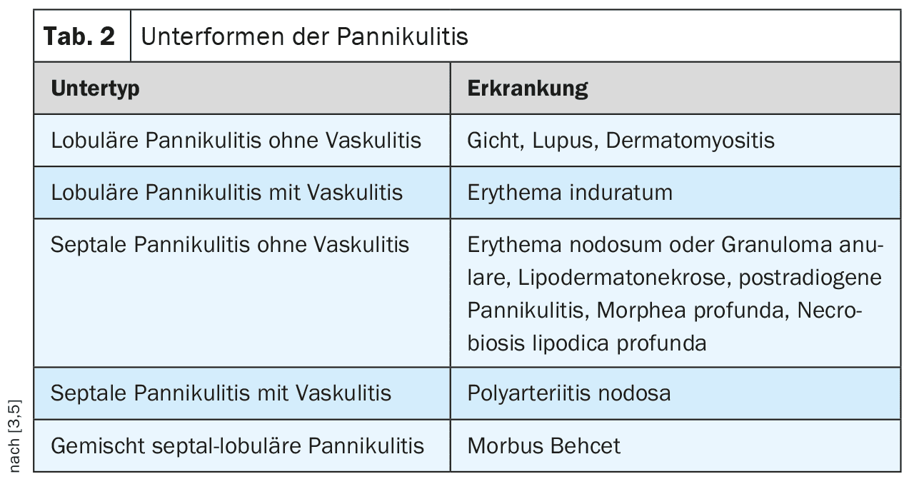
Depending on the localization of the inflammatory cells and the presence of vasculitis, panniculitis is histologically classified into different subtypes (Table 2).
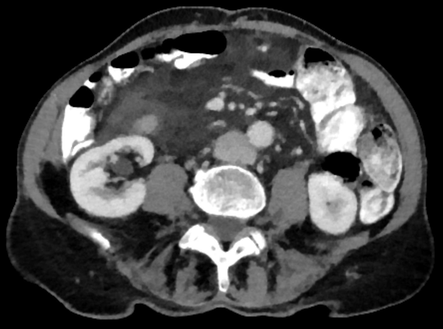
Intra-abdominal fatty tissue inflammation can also occur as mesenteric panniculitis (MP). This is a rare chronic inflammatory (or sclerosing) disease of the mesenteric fatty tissue of the small and large intestine [2].
The etiology of mesenteric panniculitis is unknown. It is more common in cancer patients, after abdominal trauma, previous abdominal surgery and obesity [1]. Table 3 lists the two main groups of MP and their characteristics.

The prevalence of MP is estimated at around 1%. It occurs in all age groups, but is more common in men in the 6. and 7th decade. The etiology is not yet fully understood, but it is classified as an IgG4-associated sclerosing disease (such as Riedel’s thyroiditis). It affects the mesenteric root of the small intestine in 90% of cases. The mesocolon or the mesoappendix are affected less frequently.
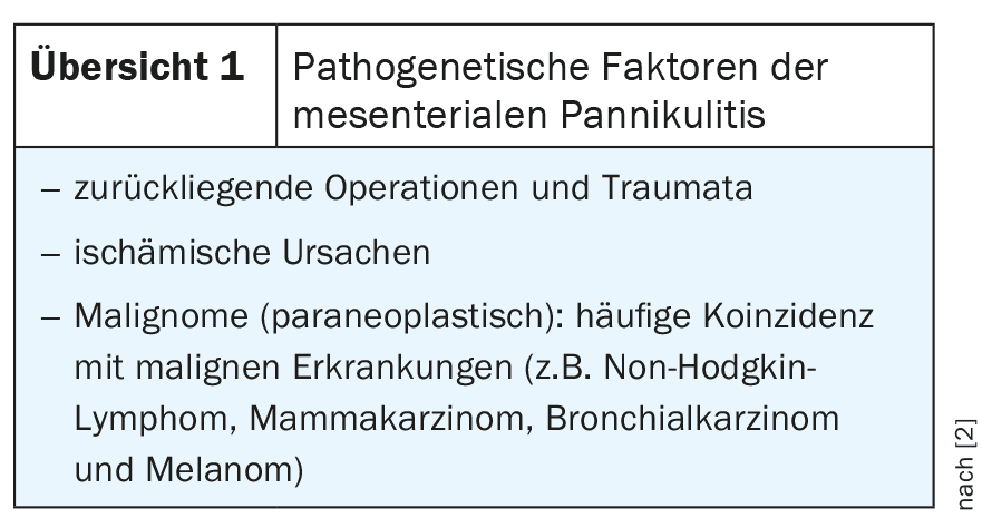
Further pathogenetic factors can be found in Overview 1.
X-rays have no significance in the diagnosis of panniculitis.
Computed tomography scans allow the inflammatory fibrous fatty tissue indurations to be well delineated [2]. Even in the native scans, connective tissue densification can be detected in the fatty tissue, which shows an increase in density in the contrast scans. Intravenous contrast is required for the detection of venous congestion syndrome.
Magnetic resonance imaging is the method of choice for visualizing inflammatory fatty tissue diseases due to its high soft tissue contrast and primary multiplanar imaging capability, especially with contrast-enhanced fat-sparing sequences.
Sonography can also show pathological changes in the fatty tissue.
Case study
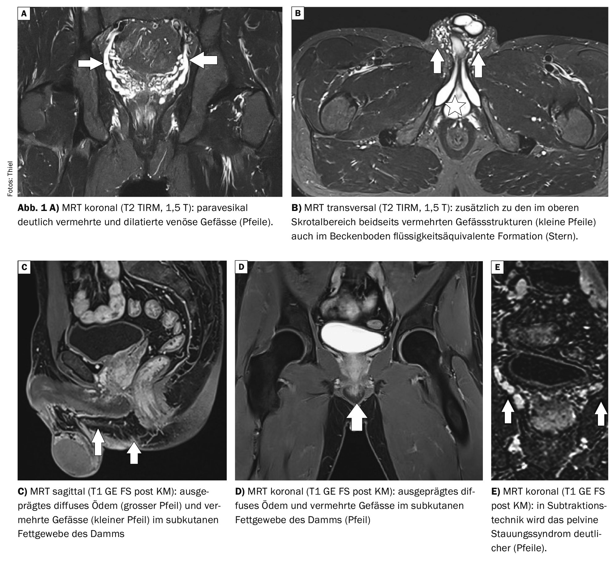
The case study demonstrates the magnetic resonance imaging diagnosis in a 38-year-old patient three months after vasectomy. About two weeks after the procedure, pain developed in the perineal area, accompanied by soft tissue swelling. External sonography described a cystoid formation in the subcutaneous fatty tissue perineally. The laboratory values were unspecific. MRI was requested to rule out an abscess or phlegmon (Fig. 1A to E). Melting inflammation could be excluded. A diffuse inflammatory reaction consistent with panniculitis was found in the subcutaneous fatty tissue. In addition, numerous dilated blood vessels were found in the pelvis and scrotum. Thus, a venous congestion syndrome was also present, analogous to pelvic congestion syndrome, which has already been presented and is characterized by dilatation of the gonadal veins and the presence of parauterine varices, occasionally also by varicosis of the vulva and venous insufficiency of the lower extremity [4].
Take-Home-Messages
- Panniculitis is a localized inflammation of the subcutaneous fatty tissue.
- It can occur in subcutaneous fatty tissue and also intra-abdominally.
- The duration of the disease varies, as do the symptoms.
- Panniculitis occurs in all age groups, predominantly in men in the 6. and 7th decade.
- Magnetic resonance imaging is the method of choice for detecting panniculitis.
Literature:
- Buragina G, Biasina AM, Carrafiello G: Clinical and radiological features of mesenteric panniculitis: a critical overview. Acta Biomed 2019 Dec 23; 90(4): 411–422.
- Decker J, et al: Mesenteric panniculitis, https://flexikon.doccheck.com,
(last accessed 11.11.2024). - Merz S, et al: Panniculitis: https: //flexikon.doccheck.com,(last accessed 11.11.2024)
- Spüntrup E, Bredel B, Steffen MS, Petzold T: Pelvic venous congestion syndrome: MR diagnostics and interventional treatment options. Obstetrics Gynecology 2022; 82(03): 269-275.
- Wick MR: Panniculitis: A summary. Semin Diagn Pathol 2017; 34(3): 261–272.
HAUSARZT PRAXIS 2024; 19(12): 42–43
Cover picture: Panniculitis mesenterialis (suspected); © hellerhoff, wikimedia