Case report: A seven-year-old girl presents with the problem of sudden “brittle” nails, some of which come off completely without pain. The symptoms started four weeks ago on single fingernails, in the course now practically all nails are affected, especially with splitting and detachment of toenails. Patient and parents are alarmed by this sudden and progressive nail problem and fear a vitamin deficiency condition or general illness. No previous skin or nail lesions are known.
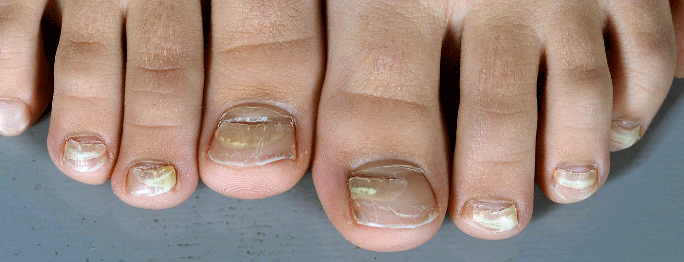
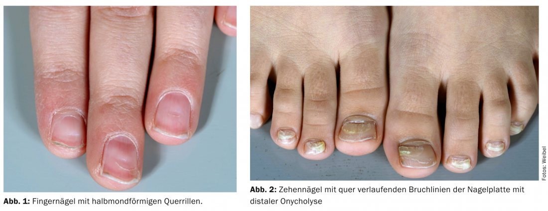
Diagnosis: On examination, most fingernails show crescent-shaped whitish transverse grooves to cracks in the center of the nail plate (Fig. 1). In comparison, the toenails show somewhat more proximal, also pronounced crescent-shaped transverse fracture lines of the nail plate with distal onycholysis and inconspicuous regrowth of the proximal nail (Fig. 2).
Quiz
What is the most common cause of this symptomatology in childhood?
AMedication intake
BKawasaki syndrome
CVaccination
DHand-mouth-foot disease
Correct answer and diagnosis: The correct answer is D: Hand-Mouth-Foot Disease (HMFK). This is acute onychomadesis and “Beau’s lines” after HMFK (Coxsackie virus infection).
Discussion: Onychomadesis describes the complete detachment of the nail plate from the matrix and occurs secondary to acute growth arrest of the nail matrix. In the sense of a milder expression, “Beau’s lines” (crescent-shaped transverse grooves) are found. The patient’s history reveals HMFK seven weeks prior to the onset of nail changes. HMFK is a relatively common coxsackie virus (enterovirus) infection in children. The course of the disease varies greatly from individual to individual, being highly febrile with disseminated small oval grayish vesicles on an erythematous base (palmoplantar, distal extremities, gluteal/genital, lips and enoral), but also accompanied by only mild general symptoms and only isolated palmoplantar vesicles. In 2000, onychomadesis was first described after HMFK in five children. Since then, several smaller local epidemics have been documented with up to >200 cases of onychomadesis after HMFK (mainly serotypes A10, A6). In the 2011-2012 North American HMFK epidemic, secondary nail changes were recorded in approximately one-quarter of cases. In Switzerland, we observe HMFK as the most common cause of acute onychomadesis in childhood. The nail changes typically occur about three to ten weeks after the viral infection.
Other typical triggers for acute onychomadesis include other high-febrile infections (noroviruses, swine flu viruses), Kawasaki syndrome, drug reactions, and acute paronychia.
Lisa Weibel, MD
Regula Wälchli, MD
Martin Theiler, MD
Literature:
- Bernier V, et al: Nail matrix arrest in the course of hand, foot and mouth disease. Eur J Pediatr 2001; 160: 649-651.
- Lopez Davia J, et al: Onychomadesis outbreak in Valencia, Spain, associated with hand, foot and mouth disease caused by enteroviruses. Pediatr Dermatol 2011; 28: 1-5.
- Mathes EF, et al: “Eczema coxsackium” and unusual cutaneous findings in an enterovirus outbreak. Pediatrics 2013; 132: e149-e157.
Dermatology Practice 2014; 24(1): 24-25