With the increasing use of imaging techniques, the incidence of incidentally detected thyroid nodules in particular has risen sharply in recent years. In addition, the thyroid gland as an important regulator of metabolic processes is increasingly becoming the focus of patient interest itself, so that more thyroid nodules are being diagnosed by targeted thyroid sonographies, e.g. as part of check-up examinations in GP practices.
With the increasing use of imaging techniques, the incidence of incidentally detected thyroid nodules in particular has risen sharply in recent years. In addition, the thyroid gland as an important regulator of metabolic processes is increasingly becoming the focus of patient interest itself, so that more thyroid nodules are being diagnosed through targeted thyroid sonographies, for example as part of check-up examinations in general practitioners’ offices.
The prevalence increases with age and also shows regional differences depending on the iodine supply. Women are affected more frequently. In two population studies, thyroid nodules were detected in the age groups >55 years in about 40-50% of men in a region in northern Germany and 65–70% in a region in southern Germany. In women, about 50–70% and 75–85%, respectively, were affected. Among those aged <35 years, thyroid nodules were much rarer (in the north <20%, in the south between about 30–40%) [1]. Iodine deficiency is considered the most important risk factor worldwide in the development of nodular goiter [2]. In Switzerland, the iodisation of table salt initiated in 1922 has been able to make the decisive contribution to the prevention of iodine-deficient goiter, with the content of potassium iodide in salt being gradually increased from 3.75 mg/kg in 1952 to 25 mg/kg as of 2014. The use of iodised table salt in private households is >80%, but the proportion in processed foods is also insufficient in Switzerland, partly as a result of the internationalisation of the industry [3].
Therefore, general thyroid ultrasound screenings are not recommended. In the workup of thyroid nodules, history, individual risk factors, and clinic should always be the primary considerations. The aim is to identify malignancies and functional autonomies in particular, but also to avoid unnecessary examinations and therapies. This article is intended to serve as a guideline on how to proceed in a structured manner after the discovery of thyroid nodules or which clarification algorithm is followed in a thyroid center and which treatment options are available.
Medical history and clinical examination
All palpable and incidentally detected thyroid nodules should be further evaluated. The first step is to take a medical history. In addition to the age of the patient, the following factors play an important role in the risk of malignancy: rapid growth, which sometimes already leads to hoarseness, previous irradiation, especially of the head/neck region, exposure to radioactive fallout (e.g., nuclear reactor events), or a family history of papillary thyroid carcinoma (i.e., at least three affected first-degree relatives). Rare overall, but representing a significant risk factor, is the familial occurrence of syndromes associated with thyroid carcinoma (e.g., multiple endocrine neoplasia type 2 (MEN2), Cowden syndrome, familial adenomatous polyposis (FAP), Carney complex [7]). During the clinical examination, the size of the thyroid gland and thyroid nodules as well as their consistency and displaceability are examined and attention is paid to asymmetry and tenderness. Likewise, it must be determined whether cervical lymphadenopathy is present. It is important to recognize whether compression symptoms, such as dysphagia, dyspnea, the already mentioned hoarseness or – very rarely – an upper influence congestion (so-called. Pemberton sign, Fig. 1) exist.

Laboratory diagnostics
This initially includes a determination of the thyroid-stimulating hormone TSH. If this is above or below the laboratory-specific reference range, the free thyroid hormones fT4 (thyroxine) and fT3 (triiodothyronine) should also be determined. The determination of fT3 is important because in focal autonomies, “T3 hyperthyroidism” may already be present while fT4 is still within the normal range. In the case of hyperthyroidism, for differential diagnostic clarification, it should be reconsidered whether, for example, iodine contamination has been present (e.g., recent administration of iodine-containing contrast medium or medication with amiodarone). In addition, the titer of the autoantibody TRAK (TSH receptor autoantibody) is very helpful. If this is elevated, it is a very specific indication of autoimmune Graves’ disease. If (subclinical) hypothyroidism is present or if corresponding parenchymal changes are seen later in the sonogram, anti-thyroperoxidase antibodies (TPO-AK) should also be measured as a sign of chronic autoimmune thyroiditis of the Hashimoto type. The determination of anti-thyroglobulin antibodies (anti-TG-AK) is not necessary and reserved for special situations such as tumor follow-up controls after differentiated thyroid carcinoma.
In thyroid nodules, the determination of calcitonin is also recommended to exclude medullary thyroid carcinoma, since ultrasound and cytology have a low specificity for this tumor entity. This leads to improved prognosis through earlier detection and has been shown to be cost-effective [8]. It should be noted that there are gender- and age-specific reference ranges and some factors may influence the value (e.g., medications such as proton pump inhibitors, renal insufficiency, etc.).
At the latest before a pending (hemi-)thyroidectomy, calcium homeostasis should also be checked to rule out primary hyperparathyroidism. Surgical repair could then be attempted in the same session, depending on the location.
Ultrasound examination
In the sonographic workup of thyroid nodules, international classification systems have been established, all of which have a very high negative predictive value to grade the risk of malignancy. However, with regard to the avoidance of unnecessary fine-needle punctures, there are differences, e.g., between the classification of the American Thyroid Association (ATA) and the European classification more commonly used in our country (EU-TIRADS), with 43.8% versus 30.7% [9]. Advantageously, the EU-TIRADS system classifies cysts and spongiform nodules with a malignancy risk of approximately 0%. Table 1 provides an overview of the ultrasound criteria.

Thyroid scintigraphy
In the presence of a decreased TSH level, thyroid scintigraphy (usually with 99mTC pertechnetate) is uncontroversial in addition to sonographic evaluation to detect focal autonomies. If, on the other hand, the TSH value is within the normal range, scintigraphy is not necessary according to the American guidelines (ATA). Nevertheless, autonomic dysfunction may be present even with normal TSH levels. This also depends on the iodine supply. Thus, in Germany, which is one of the iodine deficiency areas, scintigraphy is recommended for thyroid nodules >1 cm regardless of the TSH value. In a German multicenter study, 19% of 1262 thyroid nodules (849 patients) were diagnosed as autonomous adenomas, with TSH levels averaging 1.18 mU/l. There was an inverse correlation of the TSH level with the extent of suppression of the thyroid tissue surrounding the autonomous nodule. With complete suppression of the surrounding tissue, TSH averaged 0.42 mU/l, and with moderate suppression, TSH averaged 1.04 mU/l [10]. No precise data are available for Switzerland, which is one of the countries with a good iodine supply.
Fine needle aspiration (FNP)
In focal autonomies, the risk of malignancy is extremely low and therefore further cytologic workup is not indicated. This should definitely be taken into account, as autonomous nodules not infrequently show suspicious ultrasound features such as microcalcifications. As a consequence, misinterpretation would be frequent, since a benign follicular adenoma cannot be distinguished from a malignant follicular carcinoma. For all other thyroid nodules, including scintigraphically hypofunctional nodules, the indication for FNP is based on sonographic malignancy criteria and nodule size. Accordingly, if the risk of malignancy is considered high, the nodule should be removed from a size >10 mm to be clarified cytologically, in case of intermediate risk >15 mm and at low risk >20 mm. In the case of cysts or clearly spongiform nodules, cytological examination should be omitted (Tab. 1). If, for example, further information such as increased activity in FDG-PET/CT is available in the case of incidences of nodules, this must be taken into account in the decision. Classification systems for cytological diagnosis have also been created for standardization, on which further therapy recommendations also depend significantly. The American system used by our center (Bethesda classification with six categories, Tab. 2) is largely consistent with the British system (Thy classification with five categories) and is readily comparable. At least six groups with at least ten well-preserved and cytologically assessable thyrocytes are required for diagnostic FNP.

Molecular additional diagnostics
As mentioned above, cytologically no distinction between follicular adenomas and follicular carcinomas is possible. Also, some follicular variants of papillary thyroid carcinomas or hyperplastic nodules with follicular pattern cannot be distinguished. They are all classified as Bethesda category III (atypia of unclear significance) or IV (follicular neoplasia) and affect approximately 15-25% of all punctates [11]. The further procedure is either lobectomy for histological confirmation (in case of Bethesda IV findings) or sonographic control with repeat FNP (in case of Bethesda III findings). Particularly in the case of findings in the Bethesda III category, additional molecular genetic tests from the aspirate can therefore be helpful for more rapid decision-making. In our clinic, mutations are sought in the seven genes BRAF, KRAS, HRAS, NRAS, RET/PTC1, RET/PTC3 and PAX8/PPARG. Since only a limited number of genes are examined, the specificity and positive predictive value are high, but the negative predictive value is not sufficient. Thus, if a mutation is detected, surgical therapy is indicated. If the result is negative, FNP must be repeated, and if Bethesda III is found again, diagnostic surgery must still be recommended in the end. In the meantime, some commercial tests are also available, which examine significantly more genes, but at the same time also show losses in specificity and positive predictive value and are therefore not yet routinely recommended. Nevertheless, this area holds promise for the future and could help to retrospectively avoid unnecessary diagnostic surgery in unclear cases.
Therapy options
Thyroid surgery is recommended when thyroid carcinoma is detected (Bethesda categories V and VI), when carcinoma is highly suspected (Bethesda category IV or III with mutation detection in one of the seven genes studied), and when findings are unclear (repeated Bethesda category III without detection of a gene mutation). The extent of surgery, i.e. whether a lobectomy or total thyroidectomy is performed, depends on multifocality and the size of the carcinoma.
If medullary thyroid carcinoma has been diagnosed, total thyroidectomy is required regardless of the presence of thyroid nodules. We will not discuss special cases, such as prophylactic thyroidectomy of MEN2 gene mutation carriers, in this article.
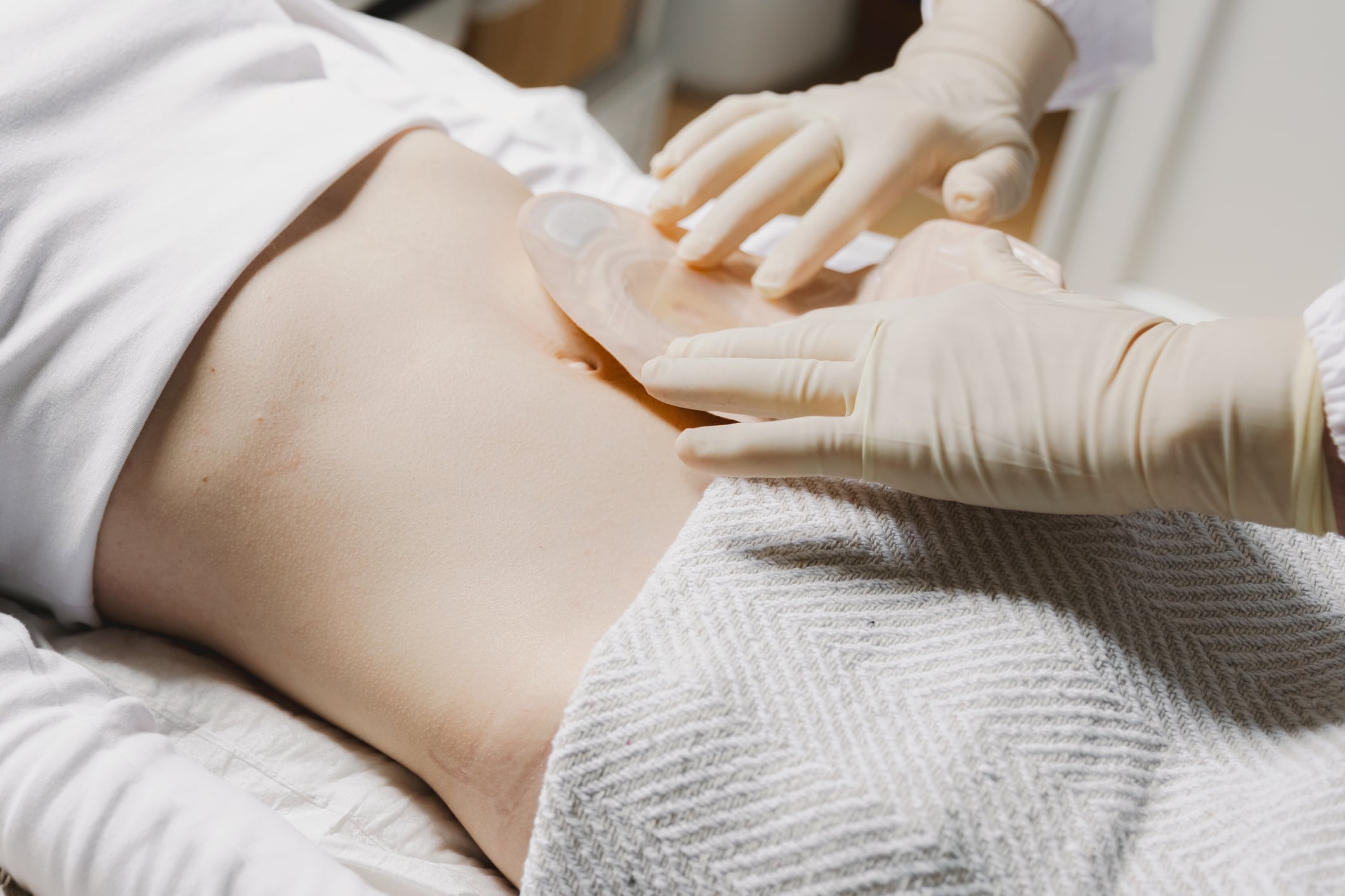
For benign thyroid nodules, surgery is the treatment of choice, especially for large multinodular strumen with compressive symptoms. If a solitary nodule causes compressive symptoms, up to a certain volume and confirmed benign lesion (FNP Bethesda category II), targeted thermal ablation of the nodule, which can be performed on an outpatient basis, can also be offered, with radiofrequency ablation (RFA, monopolar or bipolar) in particular, but also other techniques such as laser or microwave. Within 12 months, a volume reduction of up to 80% can be expected with RFA [12]. Unifocal autonomies are also a good indication for RFA (Fig. 2) . Alternatively, radioiodine therapy is a safe, non-invasive treatment option for unifocal and multifocal autonomies without compression symptoms that has been proven for decades. If autonomy is a priority, but at the same time one of the definitive therapy options is not considered – e.g. in elderly, multimorbid patients – drug therapy with a thyrostatic (carbimazole) also has its place.

In symptomatic simple thyroid cysts that do not present with complicated septation, drain well, but repeatedly refill despite two to three puncta, ethanol ablation should be discussed with the patient as a less invasive, inexpensive, and effective therapy.
Lastly, it should be mentioned that nonsuspicious asymptomatic thyroid nodules are usually followed up sonographically over a period of time, depending on the EU-TIRADS and possibly Bethesda findings. Durante et al. concluded from their prospective multicenter observation of 992 patients with 1567 thyroid nodules without additional risk factors that one year after initial diagnosis and in case of size constancy or decrease (in 85% of cases) a recheck after (3-)5 years is sufficient [13].
Take-Home Messages
- Thyroid nodules are common and should be clarified in a structured manner (clinic, laboratory, ultrasound features, scintigraphy if necessary, FNP if necessary).
- Thyroid nodules are – if necessary – well treatable. Depending on the nodal entity, there are often several options (sonographic follow-up, surgery, radioiodine therapy, thermoablation, ethanol ablation).
- Malignant thyroid nodules are rare. Treatment is curative in most cases if diagnosed in time.
Literature:
- Meisinger C, et al.: Geographic variations in the frequency of thyroid disorders and thyroid peroxidase antibodies in persons without former thyroid disease within Germany. Eur J Endocrinol 2012; 167(3): 363–371.
- Carlé A, et al.: Epidemiology of nodular goitre. Influence of iodine intake. Best Pract Res Clin Endocrinol Metab 2014; 28(4): 465–479.
- Andersson M, Herter-Aeberli I: Jodstatus in der Schweizer Bevölkerung. Schweizer Ernährungsbulletin 2018; 63–83.
- Kwong N, et al.: The Influence of Patient Age on Thyroid Nodule Formation, Multinodularity, and Thyroid Cancer Risk. J Clin Endocrinol Metab 2015; 100(12): 4434–4440.
- Haugen BR, et al.: 2015 American Thyroid Association Management Guidelines for Adult Patients with Thyroid Nodules and Differentiated Thyroid Cancer: The American Thyroid Association Guidelines Task Force on Thyroid Nodules and Differentiated Thyroid Cancer. Thyroid 2016; 26(1): 1–133.
- Vaccarella S, et al.: Worldwide Thyroid-Cancer Epidemic? The Increasing Impact of Overdiagnosis. N Engl J Med 2016; 375(7): 614–617.
- Kobaly K, et al.: Contemporary Management of Thyroid Nodules. Annu Rev Med 2022; 73: 517–528.
- Fugazzola L: Medullary thyroid cancer – An update. Best Pract Res Clin Endocrinol Metab 2022; 101655.
- Grani G, et al.: Reducing the Number of Unnecessary Thyroid Biopsies While Improving Diagnostic Accuracy: Toward the «Right» TIRADS. J Clin Endocrinol Metab 2019; 104(1): 95–102.
- Schenke SA, et al.: Distribution of Functional Status of Thyroid Nodules and Malignancy Rates of Hyperfunctioning and Hypofunctioning Thyroid Nodules in Germany. Nuklearmedizin 2022.
- Grani G, et al.: Molecular analysis of fine-needle aspiration cytology in thyroid disease: where are we? Curr Opin Otolaryngol Head Neck Surg 2021; 29(2): 107–112.
- Dobnig H, et al.: Radiofrequency ablation of thyroid nodules: «Good Clinical Practice Recommendations» for Austria. Wien Med Wochenschr 2020; 170(1–2): 6–14.
- Durante C, et al.: The natural history of benign thyroid nodules. JAMA 2015; 313(9): 926–935.
- Russ G, et al.: European Thyroid Association Guidelines for Ultrasound Malignancy Risk Stratification of Thyroid Nodules in Adults: The EU-TIRADS. Eur Thyroid J 2017; 6(5): 225–237.
- Cibas ES, Ali SZ: The 2017 Bethesda System for Reporting Thyroid Cytopathology. Thyroid 2017; 27(11): 1341–1346.
InFo ONKOLOGIE & HÄMATOLOGIE 2023; 11(4): 6–10