Medizinonline Medizinonline
  • News
    • News
    • Market & Medicine
  • Patients
    • Disease patterns
    • Diagnostics
    • Therapy
  • Partner Content
    • Diabetes
      • Dia-Month Club – Type 2 Diabetes
      • Diabetes in Motion
      • Diabetes Podcasts
    • Gastroenterology
      • IBD matters
    • Oncology
      • Swiss Oncology in motion
    • Orthopedics
      • Osteoporosis in motion
  • Departments
    • Allergology and clinical immunology
    • General Internal Medicine
    • Anesthesiology
    • Angiology
    • Surgery
    • Dermatology and venereology
    • Endocrinology and Diabetology
    • Nutrition
    • Gastroenterology and Hepatology
    • Genetics
    • Geriatrics
    • Gynecology
    • Hematology
    • Infectiology
    • Cardiology
    • Nephrology
    • Neurology
    • Emergency and intensive care medicine
    • Nuclear Medicine
    • Oncology
    • Ophthalmology
    • ORL
    • Orthopedics
    • Pediatrics
    • Pharmacology and toxicology
    • Pharmaceutical medicine
    • Phlebology
    • Physical medicine and rehabilitation
    • Pneumology
    • Prevention and health care
    • Psychiatry and psychotherapy
    • Radiology
    • Forensic Medicine
    • Rheumatology
    • Sports Medicine
    • Traumatology and trauma surgery
    • Tropical and travel medicine
    • Urology
    • Dentistry
  • CME & Congresses
    • CME continuing education
    • Congress Reports
    • Congress calendar
  • Practice
    • Noctimed
    • Practice Management
    • Jobs
    • Interviews
  • Log In
  • Register
  • My account
  • Contact
  • Publications
  • Contact
  • Deutsch
  • English
  • Français
  • Italiano
  • Português
  • Español
Subscribe
Medizinonline Medizinonline
  • News
    • News
    • Market & Medicine
  • Patienten
    • Krankheitsbilder
    • Diagnostik
    • Therapie
  • Partner Content
    • Diabetes
      • Dia-Month Club – Type 2 Diabetes
      • Diabetes in Motion
      • Diabetes Podcasts
    • Gastroenterology
      • IBD matters
    • Oncology
      • Swiss Oncology in motion
    • Orthopedics
      • Osteoporosis in motion
    • Phytotherapie
    • Rheumatology
  • Departments
    • Fachbereiche 1-13
      • Allergology and clinical immunology
      • General Internal Medicine
      • Anesthesiology
      • Angiology
      • Surgery
      • Dermatology and venereology
      • Endocrinology and Diabetology
      • Nutrition
      • Gastroenterology and Hepatology
      • Genetics
      • Geriatrics
      • Gynecology
      • Hematology
    • Fachbereiche 14-26
      • Infectiology
      • Cardiology
      • Nephrology
      • Neurology
      • Emergency and intensive care medicine
      • Nuclear Medicine
      • Oncology
      • Ophthalmology
      • ORL
      • Orthopedics
      • Pediatrics
      • Pharmacology and toxicology
      • Pharmaceutical medicine
    • Fachbereiche 26-38
      • Phlebology
      • Physical medicine and rehabilitation
      • Phytotherapy
      • Pneumology
      • Prevention and health care
      • Psychiatry and psychotherapy
      • Radiology
      • Forensic Medicine
      • Rheumatology
      • Sports Medicine
      • Traumatology and trauma surgery
      • Tropical and travel medicine
      • Urology
      • Dentistry
  • CME & Congresses
    • CME continuing education
    • Congress Reports
    • Congress calendar
  • Practice
    • Noctimed
    • Practice Management
    • Jobs
    • Interviews
Login

Sie haben noch keinen Account? Registrieren

  • Cardiomyopathy

From risk to therapy – imaging in individual risk management

    • Cardiology
    • Congress Reports
    • Radiology
    • RX
  • 2 minute read

When we talk about cardiomyopathy, we can be talking about different clinical pictures with different causes and symptoms. The actual cause may be genetic, acquired, or a mixture of both. Especially in very early or very advanced forms, the cause of the cardiomyopathy often cannot (yet) be determined. Imaging is then an important parameter to perform risk stratification of the affected individuals.

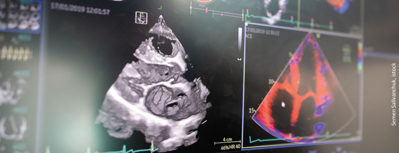
Around the question of how to stratify a patient’s risk and where to look closely, Prof. Fabian Knebel, MD, Berlin (D), explained the use of imaging as a key tool. Risk management of cardiomyopathies was addressed over one hundred years ago. At that time, the credo was still that therapy depended on the actuity of heart failure, not on its etiology. Black coffee, champagne and cognac were recommended as therapy to improve circulation. In the meantime, fortunately, we have made some progress. The current ESC position paper on cardiomyopathies recommends echocardiography as a critical imaging component after clinical findings have been obtained. This can be used to assess cardiac structure and function and to identify specific etiologies. Ultimately, dilated, restrictive, and hypertrophic cardiomyopathy should be differentiated.

Since ejection fraction (EF) alone is not sufficient, additional deformation imaging is needed. EF-based echocardiographs are guided by radial function, whereas deformation imaging can be used to capture longitudinal function. This is because patients may well have preserved pump function but reduced strain. However, these affected individuals have a much worse prognosis than patients with a normal strain, the expert said.

Targeting risk patterns

Different patterns can be detected for risk stratification. These include, for example, the hypertrophic cardiomyopathies with their genetic forms, storage diseases, neuromuscular and mitochondrial disorders, and malformations. Regarding pathomechanisms, in addition to wall thickness, we observe whether the patient has a SAM phenomenon and/or ejection tract obstruction. In addition, the gradient should be worked out. However, caution is required, especially in HCM, because different phenomena can also overlap. The strain per se forms a risk marker of HCM. If the strain is <16%, sufferers show significantly worse survival than sufferers whose strain is >16%. Mechanical dispersion can be used as a second tool for risk stratification. In addition, contrast echocardiography should be used to observe whether the septal branch into which the contrast agent was injected is the one blocking outflow.

Supportive imaging

In restrictive cardiomyopathies, the first priority is to work out by imaging whether it is a constriction with a problem primarily in the pericardium, or whether the problem is primarily in the myocardium and therefore it is a restriction. Common to both is impaired ventricular filling and diastolic heart failure. Restrictive cardiomyopathy can be divided into noninfiltrative, infiltrative disorder, storage disease, and endomyocardial disorder. These may include endomyocardial fibrosis, sarcoidosis, eosinophilic myocarditis, or Fabry disease. Specific to this patient is often that the papillary muscle is markedly thickened.


Source: risk management in cardiomyopathies. Imaging as a key tool in individual risk management. Working group meeting, 20.04.2022, 15:30-17:00
Congress: 88th Annual Meeting of the German Society of Cardiology (DGK)


CARDIOVASC 2022; 21(2): 23

Autoren
  • Leoni Burggraf
Publikation
  • CARDIOVASC
Related Topics
  • Cardiomyopathy
  • Imaging
Previous Article
  • Multiple sclerosis

The role of astrocytes in progress management.

  • Education
  • Neurology
  • Rheumatology
  • RX
  • Studies
View Post
Next Article
  • Personalized therapy in RA

Targeting instead of MTX for all

  • Congress Reports
  • Education
  • General Internal Medicine
  • Geriatrics
  • Rheumatology
  • RX
View Post
You May Also Like
View Post
  • 2 min
  • Wearables: screening via smartwatch?

New study on atrial fibrillation in high-risk patients

    • RX
    • Cardiology
    • Practice Management
    • Prevention and health care
    • Studies
View Post
  • 11 min
  • Peristomal skin health

Focus on preventive care options

    • RX
    • Cases
    • CME continuing education
    • Dermatology and venereology
    • Gastroenterology and Hepatology
    • Infectiology
    • Physical medicine and rehabilitation
    • Prevention and health care
    • Surgery
View Post
  • 5 min
  • Mental Health with GLP-1 RAs

Benefits of Semaglutide and Liraglutide

    • RX
    • Education
    • Endocrinology and Diabetology
    • General Internal Medicine
    • Nutrition
    • Psychiatry and psychotherapy
    • Studies
View Post
  • 4 min
  • Bronchiolitis obliterans

Out of shape even without popcorn

    • RX
    • Education
    • General Internal Medicine
    • Infectiology
    • Pediatrics
    • Pneumology
    • Studies
View Post
  • 8 min
  • Case study

Guillain-Barré syndrome with a fulminant course

    • RX
    • Cases
    • Education
    • Infectiology
    • Neurology
    • Pharmacology and toxicology
    • Radiology
    • Studies
View Post
  • 5 min
  • Suspicion of neuroborreliosis?

Detection of borrelia-specific CSF parameters provides certainty

    • RX
    • Congress Reports
    • Infectiology
    • Neurology
    • Studies
View Post
  • 12 min
  • The Brain and the Motivation to Eat

Why is it so hard to lose weight?

    • RX
    • CME continuing education
    • Endocrinology and Diabetology
    • General Internal Medicine
    • Nutrition
    • Pharmacology and toxicology
    • Prevention and health care
    • Psychiatry and psychotherapy
    • Studies
View Post
  • 12 min
  • Global Recommendation, Swissmedic Approval, and SL Limitation

WHO Guidelines on GLP-1 for Obesity

    • RX
    • Cardiology
    • Education
    • Endocrinology and Diabetology
    • General Internal Medicine
    • Nutrition
    • Pharmacology and toxicology
    • Prevention and health care
    • Studies
  • IBD matters

    Zum Thema
Top CME content
  • 1
    Focus on preventive care options
  • 2
    Inflammation as a Treatment Goal in Its Own Right
  • 3
    Personalized Treatment Before a Planned Pregnancy
  • 4
    Current State of Knowledge and New Therapeutic Approaches
  • 5
    Why is it so hard to lose weight?

Newsletter

Sign up and stay up to date

Subscribe
Medizinonline Medizinonline
  • Contact
  • General terms and conditions
  • Imprint

Input your search keywords and press Enter.