A young adult patient with abdominal pain was found to have hepatomegaly and splenomegaly associated with thrombocytopenia and anemia. The bone marrow was then punctured and biopsied, followed by determination of leukocyte glucocerebrosidase enzyme activity. Molecular genetic testing finally confirmed the suspected diagnosis. In the general population, the prevalence of Gaucher disease is approximately 1:100,000.
Gaucher disease is an autosomal recessive metabolic disorder due to a mutation in the glucocerebrosidase gene and a subsequent deficiency of the enzyme glucocerebrosidase. As a result, there is an accumulation of lipid-laden macrophages (so-called Gaucher cells) in the liver and spleen, as well as in the bone marrow, central nervous system (CNS), and lungs [1]. Due to the increasing overload of glucocerebrosides, the liver and spleen increase in size and displacement of the hematopoietic bone marrow occurs [2]. The consequence is, on the one hand, a reduction in platelets and erythrocytes, and on the other hand, increasing destruction of bone substance.

Case study
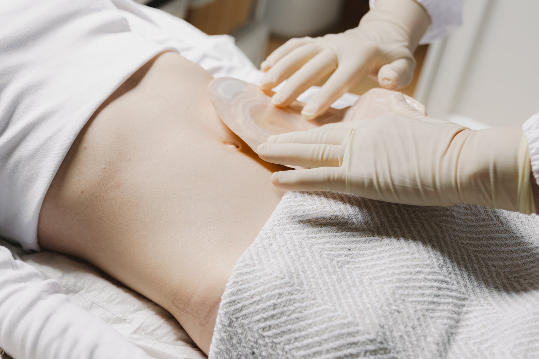
An 18-year-old female patient was admitted to the hospital with a chief complaint of right upper quadrant abdominal pain that began two weeks ago with moderate intensity and was initially slightly reduced by oral nonsteroidal anti-inflammatory drugs (NSAIDs) [3]. After a certain time, the pain-reducing effect of the NSAIDs diminished and, over time, ceased altogether. On physical examination, the conjunctiva was found to be pale and there was painful hepatomegaly 5 cm below the right costal arch. Further investigation resulted in the following findings:
- Percussion: Approximately 8 cm below the left costal arch, a dull area was noted that moved with breathing. This was the decisive factor for the patient’s admission to the hospital.
- Ultrasound examination of the abdomen: There was marked enlargement of the spleen (20.1 × 8 cm), without nodules; the liver was more than 6 cm below the costal margin, without nodules; no enlarged abdominal lymph nodes; small amount of free fluid in the peritoneal cavity adjacent to bowel loops of normal diameter; other organs without changes.
- Bone marrow aspiration: hypercellular bone marrow, with megakaryopoietic and erythropoietic hyperplasia; granulopoietic integrity. Increase in eosinophil precursors; focal increase in mature, well-differentiated lymphocytes; increase in plasma cells less than 10%; abundant histiocytes with clear cytoplasm resembling Gaucher cells, without evidence of hemophagocytosis.
- Bone marrow biopsy: Bone marrow cylinders with more than 6 marrow spaces with histiocytic cells that had abundant clear cytoplasm in vacuoles masked by remaining hematopoietic tissue. The appearance is suggestive of lysosomal storage disease.
Following the biopsy result, glucocerebrosidase enzyme activity was determined in peripheral leukocytes. This was 0.020 nmol/mg/h (normal value: 0.12 nmol/mg/h). Subsequent molecular genetic testing finally revealed double heterozygosity for the L444P and N370S mutations , confirming the diagnosis of Gaucher disease.
| Important facts about M. Gaucher at a glance Gaucher disease belongs to the group of lysosomal storage diseases. Since the individual symptoms can also have other causes, diagnosis often proves difficult. The clinical picture was first described in 1882 by Philippe Charles Gaucher, who reported a patient with splenomegaly. The following three clinical phenotypes of Gaucher’s disease are distinguished [2]: Type 1: non-neuropathic form, Type 2: acute neuropathic form, Type 3: chronic neuropathic form. Adults manifest primarily type 1 or 3. Symptoms typical of Gaucher’s disease patients include bone pain, a decrease in performance, and a tendency to bleed. Furthermore, Gaucher’s disease is associated with an increased susceptibility to infections [4]. Laboratory diagnostics typically reveal anemia and thrombocytopenia. The imaging findings are groundbreaking: ultrasound reveals enlargement of the liver and spleen, and X-rays or MRIs show bone changes with destruction of the baelcae structure as well as bone infarcts, which are initially visible in the long tubular bones of the legs. Rarely, pulmonary hypertension is found due to incorporation of glucocerebrosides into the macrophages of the lungs. In cases of clinical suspicion of Gaucher disease and corresponding laboratory and imaging findings, it is advisable to determine the enzyme activity of glucocerebrosidase in a specialized laboratory. Nowadays, enzyme replacement therapy with recombinant human glucocerebrosidase or substrate reduction therapy are available as treatment approaches [2,5,6]. |
Literature:
- Grabowski GA: Phenotype, diagnosis, and treatment of Gaucher’s disease. Lancet 2008; 372: 1263-1271.
- Tran C, et al: Pulmonary involvement in adult patients with inborn errors of metabolism. Compass Pneumol 2018; 6 (1): 6-17.
- Valdés-Díaz K, et al: Gaucher disease. Presentation of a clinical case and literature review. Hematol Transfus Cell Ther 2022; 44(1): 104-107.
- Gaucher’s disease, www.uniklinik-duesseldorf.de/patienten-besucher/klinikeninstitutezentren/klinik-fuer-gastroenterologie-hepatologie-und-infektiologie/klinik/fuer-patienten/behandlungsschwerpunkte/stoffwechselkrankheiten/morbus-gaucher,(last accessed Sept. 18, 2023).
- Goitein O, et al: Lung involvement and enzyme replacement therapy in Gaucher’s disease. QJM 2001; 94: 407-415.
- Shemesh E, et al: Enzyme replacement and substrate reduction therapy for Gaucher disease. Cochrane Database Syst Rev 2015; 3: CD010324.
HAUSARZT PRAXIS 2023; 18(9): 26