Flegel’s disease is a genodermatosis with manifestation in the second half of life. Topical topical preparations such as those containing urea or cortisone are currently used for therapy. To clarify the genetic cause of this rare dyskeratosis, a research team from Germany and Switzerland conducted a study using next-generation sequencing. The results of the analysis provide valuable insights for the development of new therapeutic approaches.
In 1958, the German dermatologist Heinz Flegel was the first to describe a new, rare skin disease characterized by multiple, asymptomatic, hyperkeratotic, reddish-brown flat papules on the extensor sides of the extremities, especially in the lower extremities and dorsum of the foot [1]. The clinical picture is called hyperkeratosis lenticularis perstans (HLP) or Flegel’s disease. Typically, the skin symptoms first appear in the fourth to fifth decade of life. Regarding etiology of HLP, there are still many open questions. A genetic cause has been suspected for some time, but so far no gene variants have been clearly attributed to HLP.
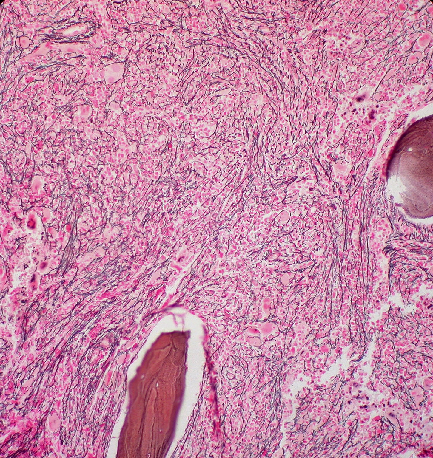
| Case vignette A 72-year-old Caucasian woman presented for multiple small, reddish and brownish, keratotic papules. The papules first appeared on the lower limbs and later spread to the upper limbs and trunk (Fig. 1A and B). There was no history of previous cutaneous or systemic disease, except for an in situ well-differentiated cutaneous squamous cell carcinoma of the leg that had been removed two years earlier. The dermoscopy findings are shown in Figure 2. Histopathology of a lesion on the trunk showed compact, thick lamellar hyperkeratosis with focal parakeratosis over the atrophic epidermis associated with a dense, lichenoid infiltrate of small lymphocytes in the superficial dermis with focal accentuation in the papillomatous areas. Immunohistochemical studies revealed a similar proportion of CD4 and CD8 T lymphocytes accompanied by sparse to absent CD20 cells. After clinicopathologic correlation, a final diagnosis of generalized hyperkeratosis lenticularis perstans (Flegel’s disease) was made. |

“Loss-of-function” variants identified in the SPTLC1 gene.
To elucidate the genetic cause of HLP, researchers retrospectively analyzed EDTA** blood, skin biopsies, and formalin-fixed paraffin-embedded (FFPE) tissue from five patients using next-generation sequencing (NGS) [1,2]. Different rare heterozygous germline variants in the gene SPTLC1 (Serine Palmitoyltransferase Long Chain Base Subunit 1) could be identified in all patients. Interestingly, these loss-of-function variants were either small deletions or splice variants. Indeed, immunofluorescence staining of skin sections showed reduced SPTLC1 staining in HLP patients.
** Blood that has been unclotted with the chelating agent ethylenediaminetetraacetate (EDTA) for further examination in laboratory medicine.

In addition, there was evidence of a “second hit” in affected lesions in the form of a “loss of heterozygosity”. Based on these results, the study authors concluded that pathogenic germline variants in the SPTLC1 gene in combination with a “loss of heterozygosity” in lesional skin are responsible for the development of HLP. This likely results in a decrease in SPTLC1 protein – a key enzyme in sphingolipid biosynthesis – and subsequent disruption of the sphingolipid signaling pathway. The elucidation of the genetic causes of Flegel’s disease is an important basis for the development of new therapeutic approaches for this rare skin disease, according to the authors [1,2].
Literature:
- Jägle S, et al: Pathogenic variants in the SPTLC1 gene are causative for Flegel’s disease (hyperkeratosis lenticularis perstans). FV02/10th DDG Meeting, 26-29.04.2023
- Jägle S, et al: Pathogenic variants in the SPTLC1 gene cause hyperkeratosis lenticularis perstans. Br J Dermatol 2023; 188(1): 94-99.
- Stabile G, et al: Widespread and Long-Enduring Hyperkeratosis Lenticularis Perstans (Flegel’s Disease): Clinico-Pathological and Dermoscopic Features of a Rare Presentation. Dermatopathology 2023; 10(1): 46-51. www.mdpi.com/2296-3529/10/1/6,(last accessed Aug 18, 2023).
DERMATOLOGY PRACTICE 2023; 33(4): 40