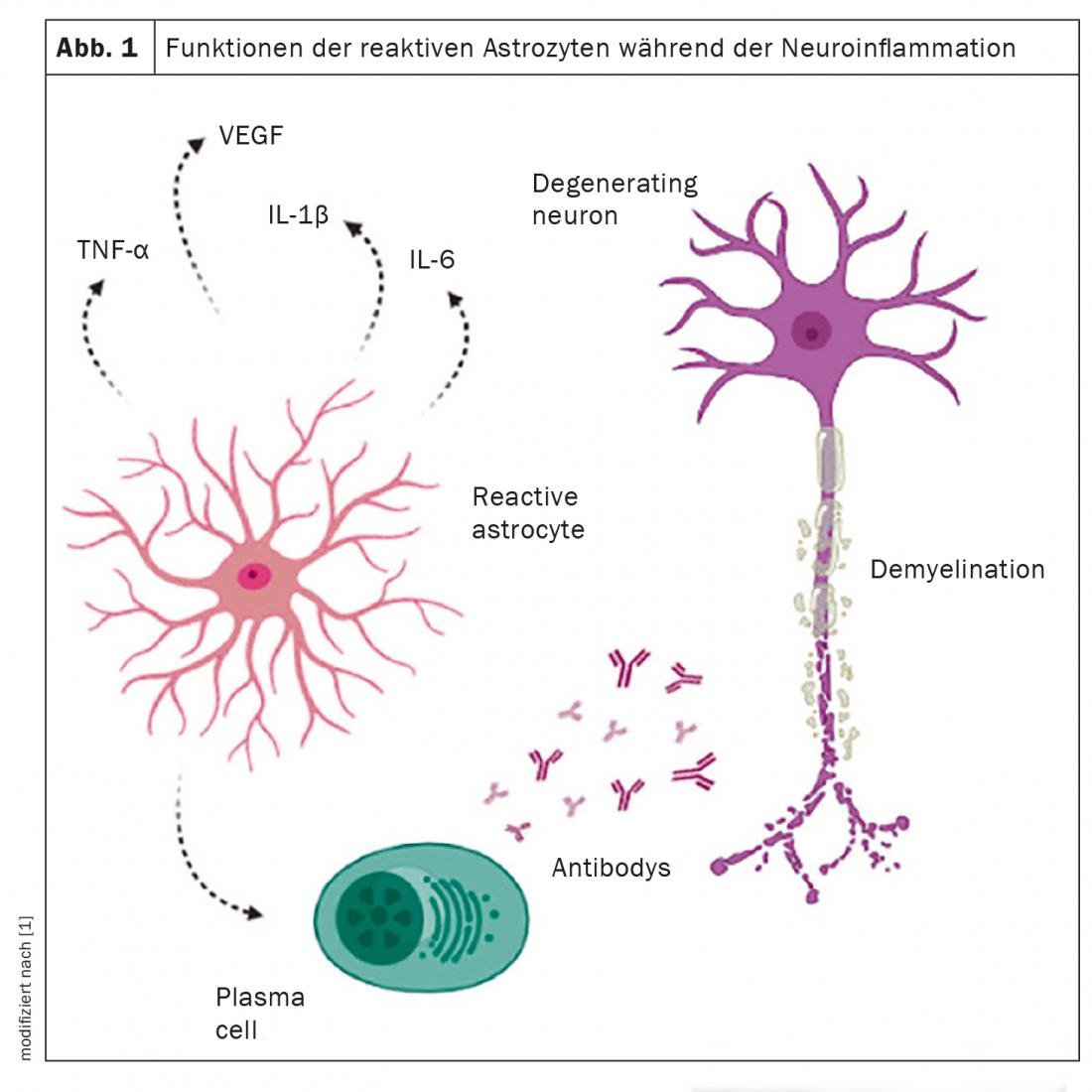
In chronic neurodegenerative diseases such as multiple sclerosis, CNS cells make various adaptations during neuroinflammation. The main cells involved in this inflammatory process are the glial cells, among which the astrocytes stand out. Reactive astrocytes have been reported to lose their supportive function and take on a toxic function with disease progression.
Astrocytes represent the most abundant cell group in all regions of the central nervous system. Depending on the type of lesion, they may secrete both pro-inflammatory cytokines (Th1, IL-1, IL-2, IL-6, IL-7, and TNF) and anti-inflammatory ones (Th2, IL-4, IL-10, IL-13, and TGFβ). In addition, they are associated with several physiological functions, including nutrient secretion, maintenance of the neuronal microenvironment, blood-brain barrier permeability, and development of pathological processes in the brain. Astrocytes play a complex role in the pathogenesis of neurodegenerative diseases such as multiple sclerosis (MS). Several studies report that reactive astrocytes lose their supportive role and acquire a toxic function as these diseases progress. A review therefore highlighted the key features of astrocytes in the development of MS to provide a better understanding of the disease.
The secret stars of the brain
Astrocytes belong to the glial cells, i.e. the non-electrically excitable cells of the nervous system. They are composed of many intertwined fibrils, the structure of which is called a glial filament. They can be divided into two main subtypes, termed fibrous or protoplasmic, based on differences in their cell morphology and glial filament content. Fibrous astrocytes are mainly located in the white matter and have long fibers with many glial filaments in the cytoplasm. Protoplasmic astrocytes, in turn, are widely distributed in the gray matter and exhibit thick branching. The formation of neurovascular units occurs through astrocytic processes, as these cells act as a bridge between neurons and blood vessels. In addition, astrocytic cells can provide structures and metabolic support for neurons and play important roles in regulating neuronal survival, synapse formation, and ion channel distribution.
Astrocytic podocytes closely surround endothelial cells and are critical to the structure of the blood-brain barrier because of their ability to form tight junctions and their high mitochondrial content. Through their interactions with components of the blood-brain barrier (BBB), astrocytes not only regulate its function but also recognize molecules produced by peripheral immune cells, including cytokines. They also express numerous receptors that enable them to respond to neuroactive compounds such as neurotransmitters, neuropeptides, growth factors, cytokines, and toxins.
Their role in the MS disease process
The main pathological feature of MS is the presence of focal inflammatory lesions and demyelination caused by the immune response. The role that astrocytes play in the development of these lesions is active and diverse, with several functional changes, such as altering the permeability of the blood-brain barrier, which favors a deregulated immune response to the central nervous system.
Astrocytes respond to CNS injury with a complex activation process that includes morphological, transcriptional, and biochemical changes as well as functional changes associated with a reduction in homeostatic metabolic functions. This is accompanied by increased expression of the glial fibril intermediate filament (GFAP) acidic protein, which therefore serves as a marker for reactive astrocytes. Because they respond to changes in their environment, it is difficult to distinguish between normal cell and reactive astrocyte. A reactive astrocyte is the cell that has responded to a disease or tissue change. Astrocytes are present at the active margins of demyelinating lesions and play a key role in removing damaged myelin. These cells are hypertrophic, have large nuclei, and their cytoplasm may contain glial intermediate filaments and cellular debris. In chronic lesions, the astrocytic peripheral rim shows active demyelination and may cause further inflammatory damage that contributes to disease progression.
However, astrocytes play a dual role: they not only contribute to axonal degeneration and demyelination, but also create a favorable environment that promotes remyelination. However, the influence of astrocytes on the pathogenesis and repair of the inflammatory process depends on several factors, such as the timing after injury, the type of injury, the surrounding microenvironment, the interaction with other cell types, and factors affecting their activation. The various neurotrophic factors, cytokines, and growth factors provided by astrocytes facilitate the important repair process in MS. Furthermore, astrocytes further support remyelination by recruiting macrophages/microglia, which are responsible for removing myelin remnants from the sites of demyelinated injury, thereby enabling the formation of new myelin.
Astrocytes as a therapeutic target
Given the importance of astrocytes in the pathogenesis of MS, they offer an attractive therapeutic target. None of the currently approved treatment options specifically address astrocytes. However, several therapies have shown an effect on them. Dimethyl fumarate (DMF) inhibits proinflammatory activation of astrocytes, including NF-κB signaling, and activates the transcription factor Nrf2, which regulates the antioxidant response in astrocytes. Fingolimod acts on astrocytes by inhibiting NF-κB signaling, reducing the expression of pro-inflammatory cytokines, and increasing neurotrophic factors. Laquinimod reduces NF-κB signaling and astrocyte pro-inflammatory responses. Glatiramer acetate (GA) induces IL-10 and transforming growth factor (TGF-β) expression by astrocytes, restores perivascular astrocyte connections with blood vessels and neuronal synapses, and inhibits TNF-α.
Source:
- Salles D, Spindola Samartini R, de Seixas Alves MT, et al: Functions of astrocytes in multiple sclerosis: A review. Multiple Sclerosis and Related Disorders 2022; 60: 103749.
Further reading:
- Aharoni R, Eilam R, Arnon R, et al: Astrocytes in multiple sclerosis-essential constituents with diverse multifaceted functions. Int J Mol Sci 2021; 22: 11.
- Ambrosini E, Remoli ME, Giacomini E, et al: Astrocytes produce dendritic cell-attracting chemokines in vitro and in multiple sclerosis lesions. J Neuropathol Exp. Neurol 2005; 64(8): 706-715.
- Batiuk MY, Martirosyan A, Wahis J, et al: Identification of region-specific astrocyte subtypes at single cell resolution. Nat Commun 2020; 11: 1220.
InFo NEUROLOGY & PSYCHIATRY 2022; 20(3): 35-35.