Diseases of the spinal cord are often accompanied by a great reduction in the quality of life for those affected. Cervical spondylotic myelopathy is the most common cause of myelopathy in older age. Differential diagnoses of cervical spondylotic myelopathy to consider include the entire spectrum of myelopathies. Close interdisciplinary collaboration between neurologists, neurosurgeons, and orthopedists is required when deciding between a conservative vs. surgical approach. Case-by-case decisions must be made, as controlled studies on this therapeutic decision are lacking.
The spinal cord, as part of the central nervous system, is the continuation of the medulla oblongata and in adults extends from the foramen magnum down to the first or second lumbar lumbar vertebra. It is surrounded and well protected by the bones and ligaments of the spinal canal. However, depending on the anatomy and pathological alteration, this actually protective spinal canal itself may pose a threat to the spinal cord by causing spinal stenosis with consecutive cervical spondylotic myelopathy (CSM) [1].
CSM is a condition that must be addressed in close interdisciplinary collaboration between neurologists, neurosurgeons, and orthopedic surgeons. This close collaboration is important in that no general approach is possible due to the individuality and complexity of the disease, and controlled studies on this topic are lacking. Therefore, the consensus building of the mentioned specialties must always be strived for a goal-oriented treatment.
Definition
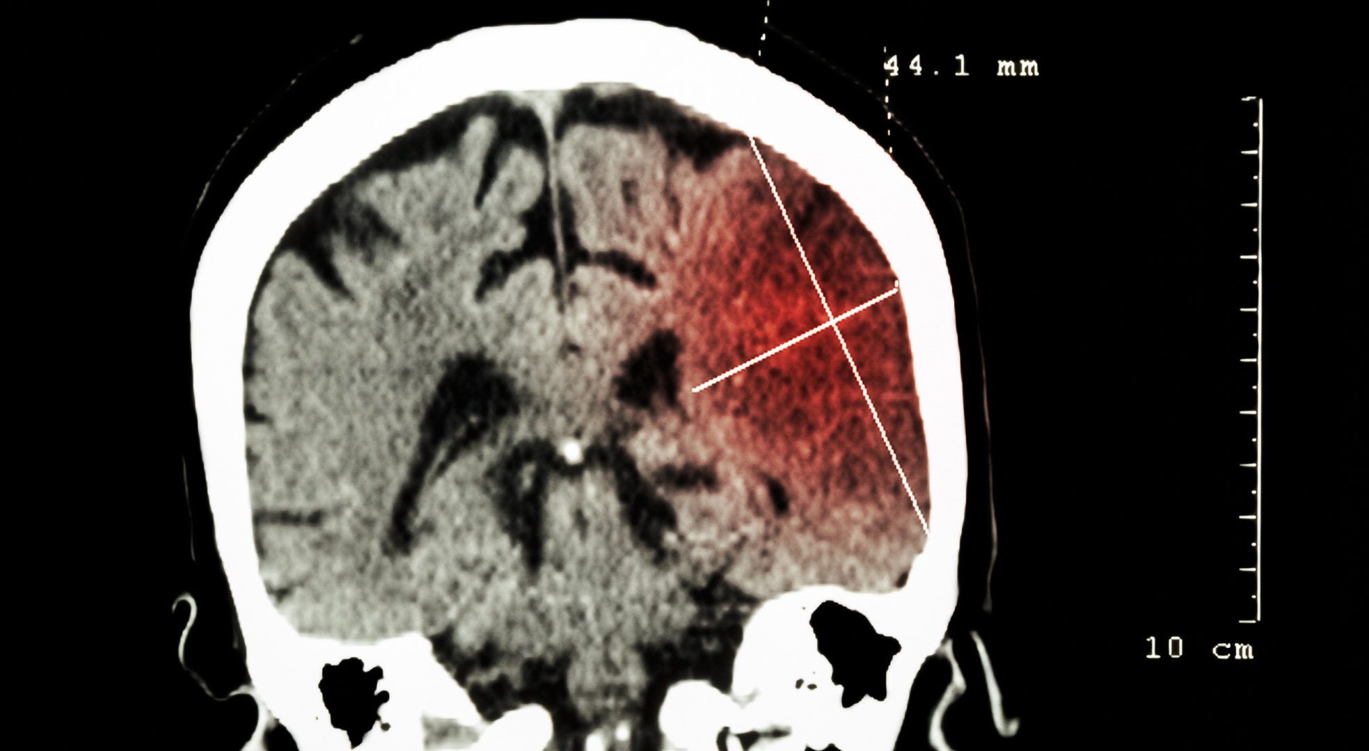
CSM is an age-related disease of the cervical spine that can cause damage to the spinal cord and nerve roots due to compressive changes. CSM is the most common cause of myelopathy in older age. Interestingly, a high percentage of the elderly have degenerative changes of the cervical spine detectable by imaging morphology, but by far not all develop CSM. Typically, progressive degenerative changes must occur on the floor of a preexisting spinal stenosis for a clinical manifestation of CSM to occur (Fig. 1).

Pathophysiology
The underlying pathophysiology of CSM is not always clear. The resulting pressure on the spinal cord and nerve roots leads initially to damage of the myelin sheaths, but in the course also to an axonal lesion. Depending on the level of the lesion and the neuronal structure affected, the corresponding clinical pattern of failure occurs, but also pain as a warning symptom. In addition to these purely compression-related causes, vascular problems also appear to worsen the situation. A reduction in arterial blood supply or venous outflow may be followed by spinal cord edema, with a corresponding progression of myelopathy. Inflammatory and biomolecular factors also play a role [2]. Typically, it is a triad of spinal cord compression, vascular compression with consecutive ischemia, and the appearance of intramedullary edema, which together drive the problem of myelopathy [1].
Clinic
According to the damage to the spinal cord and the nerve roots involved, a multifaceted neurological pattern of deficits is seen. The symptoms range from mild paresthesia of the fingers bds. on signs of a lesion of the first motor neuron with pyramidal tract signs in the sense of spastic muscle tone elevation and increased muscle reflexes. Due to the afferent disorder in affected sensitive structures, a broad-based, ataxic gait pattern is seen. Disorders of bladder-rectum function may occur. In the area of the directly damaged cervical nerve roots, the clinical-neurological picture is one of a peripheral deficit syndrome with atrophic paresis of the corresponding characteristic muscles accompanied by radicular sensory disturbances and pain as well as possible weakening of the muscle reflexes at the level of the lesion [3].
Differential diagnosis
Differential diagnosis of cervical canal stenosis must always be considered in cases of slowly progressing paraparesis, posterior cord ataxia or gait disturbance, especially in older age. Of course, neurologic symptoms may also develop subacutely or acutely when the cervical canal has fallen below a critical narrowing or by compression of the vessels. At the level of the cervical stenosis, there may be radicular pain that mimics the symptoms of a herniated disc. Disc herniation is the most common cause of a space-occupying lesion of the spinal cord; other possible space-occupying lesions include epidural or intradural tumors such as meningiomas (Fig. 2), cavernomas (Fig. 3), or an epidural abscess. Traumatic spinal injuries can also lead to myelopathy (Fig. 4).


Overall, the differential diagnosis of a CSM encompasses the entire spectrum of myelopathies [3]. From a nosologic standpoint, myelopathy is a broad term analogous to “neuropathy” or “encephalopathy,” which may be based on traumatic, autoimmune, infectious, neoplastic, vascular, or hereditary degenerative disease of the spinal cord. Examples of hereditary degenerative spinal cord diseases are hereditary spastic spinal paralysis, spinal muscular atrophy, Friedreich’s ataxia, or the various courses of spinocerebellar ataxias (SCA).
Depending on the level of damage and the extent of the lesion, a cross-sectional picture with paresis accompanied by sensory disturbances and bladder-rectum disturbances is seen. Lesions of the superior cervical spinal canal predispose to the occurrence of tetraparesis, and lesions of the inferior cervical and thoracic regions lead to paraplegia of the legs. It must always be kept in mind when evaluating neuroradiologic imaging that the morphologic lesion may be above the clinically graspable level.
Rarely, transverse myelitis can also mimic the picture of cervical canal stenosis, again resulting in progressive paraparesis of the legs, including loss of sensation and disturbances in sphincter function. However, the course of myelitis is generally more rapid than that of osseous spinal stenosis, and patients are usually younger. Often the complaints are associated with pain.
Furthermore, in the case of acute clinical symptoms, spinal ischemia in the supply area of the anterior spinal artery should also be considered, which leads to damage of the descending pyramidal tract fibers with paraparesis below the ischemic area. Lesion of the lateral spinothalamic tract may result in loss of pain and temperature sensation. At the same time, the backbone function remains intact in terms of surface sensitivity. Due to this constellation with preserved touch sensation, the diagnosis of a psychogenic disease is not infrequently inaccurately made. Spinal anterior syndrome may occur intermittently with recurrent paresis of the legs due to transient ischemic attacks in the anterior spinal artery supply area. This is seen in terms of vascular spinal claudication, but also in underlying arteriovenous spinal fistulas (Fig. 5).

The main spinal symptoms, their anatomical assignment and consequent clinical manifestation are summarized in Table 1.

Diagnostics: Additional technical examinations and laboratory
The first step in the diagnostic process is to take a careful medical history and to determine the clinical-neurological findings. In particular, a possible cervical spinal stenosis must be considered at all, which is often overlooked in clinical practice. Regarding neuroradiologic imaging, an MRI scan is particularly useful for targeting [4–6].
The most sensitive MRI sequences to detect spinal cord lesions are the STIR (“short-tau inversion recovery fast spin-echo”) sequence and the T2-weighted “fast spin” echo sequence [3–5]. In individual cases, there may also be an indication for an additional MRI examination of the skull with the question of the presence of a subcortical vascular encephalopathy or in order to be able to exclude an inflammatory CNS process that goes beyond a myelopathy. Electrophysiologic diagnostics such as electroneurography, electromyography, or somatosensory evoked potentials (SSEP) are often helpful, especially to rule out peripheral nervous system pathology such as immune-mediated neuropathies, which can also mimic the presence of myelopathy. Motor evoked potentials (MEP) can be helpful to differentiate between motor neuron disease such as amyotrophic lateral sclerosis and cervical spondylotic myelopathy by using the triple stimulation technique [7].
If transverse myelitis is suspected, CSF diagnostics must be performed, including immunoglobulin index (Reiber scheme), oligoclonal band determination, and cytologic analysis. Vitamin B12, methylmalonic acid, HIV screening, Borrelia and syphillis serology, thyroid hormones, and Vit D3 levels should also be determined if transverse myelitis is suspected. Table 2 summarizes the differential diagnoses of transverse myelitis [8].

Therapy
Many studies have shown that surgical therapy leads to sustained improvement over the long-term. Unfortunately, however, these studies did not directly compare a surgical versus a conservative approach [2]. The question of primarily conservative or surgical therapy must therefore be answered taking into account the overall situation. This takes into account the severity and duration of the clinically tangible symptoms, the time course and also the patient’s age. The results of neuroradiological diagnostics are important for the therapeutic decision, but the indication is mainly based on the clinical picture. A conservative approach is reasonable if the patient has few neurologic deficits and there is no radiological evidence of myelopathy. In this case, however, in addition to a clinical neurological follow-up examination, a follow-up MRI examination must also be performed after three to six months. In any case, regular physiotherapy on a neurophysiological basis is particularly important, with the aim of building up muscles and thereby stabilizing the cervical spine. Supportive complementary medicine procedures can be helpful for affected individuals [9]. Inpatient neurologic rehabilitation therapy may also be appropriate if a decision is made to proceed conservatively. Temporary antiphlogistics and possibly also muscle relaxants may be considered.
The indication for surgical rehabilitation must be examined particularly carefully. Especially in cases of long-standing and clinically neurologically pronounced symptoms, the prognosis regarding postoperative improvement is difficult to predict. Additional electrophysiologic diagnostics can aid in decision making in addition to image morphology on MRI. It is important to note that the clinically neurologically graspable findings must correlate with the image morphologic features and this is appropriately supported by the electrophysiologically graspable pattern of failure [10].
Acute to subacute paraplegic symptomatology that develops rapidly and is due to demonstrable CSM after exclusion of other possible causes of myelopathy is an indication for surgery. A legitimate goal of surgery may also be to prevent the progression of gait dysfunction, fine motor dysfunction of the hands, or bladder dysfunction.
Once the decision has been made to surgically repair the spinal stenosis, the choice of surgical technique (ventral vs. posterior procedure) and the extent of the procedure is left to the specialists in the surgical disciplines. In the case of bladder-rectum dysfunction, neuro-urological interdisciplinary cooperation is required.
Acknowledgments: My sincere thanks go to Dr. Johannes Weber, Senior Physician, Department of Radiology, Cantonal Hospital St. Gallen, for selecting and providing the illustrations.
Literature:
- Beattie MS, Manley GT: Tight squeeze, slow burn: inflammation and the etiology of cervical myelopathy. Brain 2011; 134: 1259-1263.
- Wilson JR, et al: State of the Art in Degenerative Cervical Myelopathy: An Update on Current Clinical Evidence. Neurosurgery 2017 Mar 1; 80(3S): S33-S45.
- Tettenborn B, Hägele-Link S.: Spinal Cord Disorders. Eur Neurol 2015; 74(3-4): 141-146.
- Bot JC, et al: Comparison of a conventional cardiac-triggered dual spin-echo and a fast STIR sequence in detection of spinal cord lesions in multiple sclerosis. Eur Radiol 2000;10(5): 753-758.
- Campi A, Pontesilli S, Gerevini S: Comparison of MRI pulse sequences for investigation of lesions of the cervical spinal cord. Neuroradiology 2000; 42(9): 669-675.
- Wheeler-Kingshott CA, et al: The current state-of-the-art of spinal cord imaging: Applications. Neuroimage 2014; 84: 1082-1093.
- Truffert A, Rösler KM, Magistris MR: Amyotrophic lateral sclerosis versus cervical spondylotic myelopathy: a study using transcranial magnetic stimulation with recordings from the trapezius and limb muscles. Clin Neurophysiol 2000 Jun; 111(6): 1031-1038.
- Tettenborn, B: Paroxysmal paresis. In: Tettenborn B, Schmitz B (eds.): The paroxysmal disorders. Cambridge University Press 2010; 89-112.
- Ostermann T, et al: Effects of Rhythmic Embrocation Therapy With Solum Oil in Chronic Pain Patients: A Prospective Observational Study. Clin J Pain 2008; 24: 237-243.
- Debette S, de Seze J, Pruvo JP: Long-term outcome of acute and subacute myelopathies. J Neurol 2009; 256: 980-988.
InFo NEUROLOGY & PSYCHIATRY 2017; 15(3): 14-18.