Light urticaria (urticaria solaris) is a rare photodermatosis whose diagnosis and treatment have not yet been standardized. The results of a long-term study were published in one of this year’s first issues of Acta Dermato-Venereologica. Pesqué et al. aimed to analyze clinical and therapeutic characteristics in a cohort of patients with light urticaria in long-term follow-up, with a focus on treatment courses with antihistamines and omalizumab.
The study was conducted between January 2007 and May 2023 at a department specializing in urticaria at the Hospital del Mar, Barcelona (Spain) in adult patients diagnosed with light urticaria (Box) [1]. The diagnosis was based on anamnestic data and photoprovocations, which were used to determine the range of the triggering wavelengths. A total of 41 patients were diagnosed with light urticaria during this period. The mean onset of the disease was 34.0 years. The gender ratio was dominated by women (n=26; 63.4%). Contact allergic dermatitis, atopic dermatitis and other subtypes of chronic inducible urticaria (CindU) were common comorbidities, while only a minority of light urticaria patients had chronic spontaneous urticaria (CSU) or autoimmune diseases (less than 5% each).
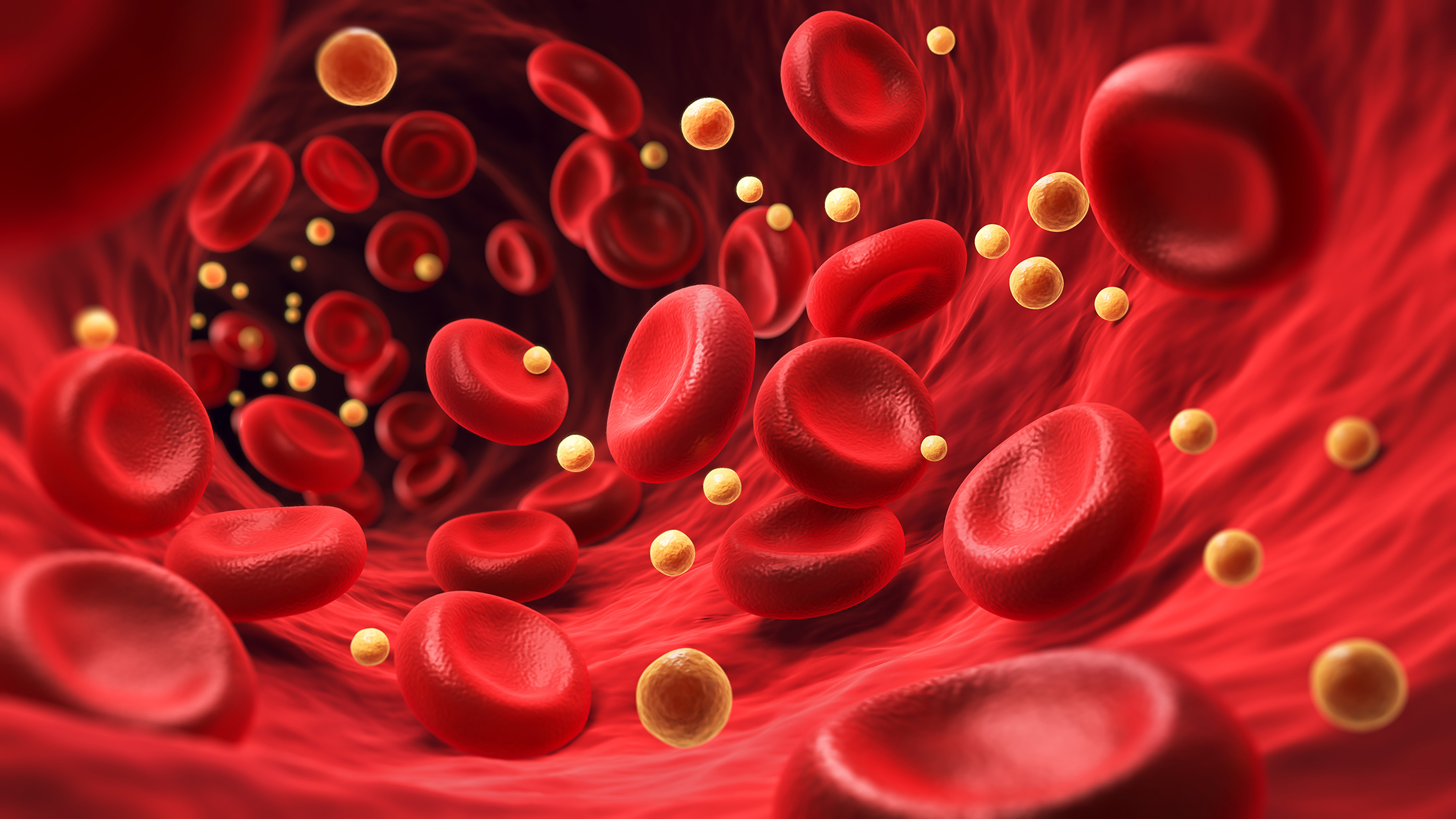
| Although the exact pathogenesis and etiology of light urticaria are not yet fully understood, it is assumed that, similar to an immediate-type allergic reaction (type I according to Coombs and Gell), specific immunoglobulin E (IgE) antibodies, which are triggered by photoallergens, bind mast cells, leading to the formation of wheals [1–4]. The skin reactions caused by ultraviolet radiation (UVA and/or UVB) and/or visible light in the context of light urticaria occur within a few minutes of exposure and begin with pronounced itching, followed by erythema and wheal formation of varying intensity depending on the dose [2]. Severe cases with headaches, dizziness, nausea, bronchospasm, arterial hypotension and tachycardia up to anaphylactic shock are also possible, particularly if large areas of skin are exposed to the triggering light spectrum [2,5]. |
Light urticaria episodes were accompanied by hives (n=41, 10%), itching (n=39, 95.1%), burning (n=11, 26.8%) or even angioedema (n=3; 7.3%). No associated systemic symptoms were observed. The skin symptoms occurred within a median of 5 minutes after exposure and lasted a median of 60 minutes. Most patients responded positively to photoprovocation (n=38; 92.7%), with the most common range of triggering wavelengths being the combination of UVA and visible light (n=11; 29.7%).
Antihistamines as first-line treatment are mostly effective
The mean baseline IgE serum level was 136.5 UI/ml (IQR 338.5-50.5 UI/ml) [1]. Autoantibodies (ANA and anti-TPO) were only detected in a small proportion of patients (9.8%). The findings regarding porphyrins were unremarkable. The median follow-up time of the cohort was 60.0 months. The vast majority of patients (n=40; 97.6%) used photoprotection. Antihistamines were the treatment of first choice in all patients; initially at a 2-fold dose and if this was not sufficient to achieve control, a 4-fold dose was prescribed (n=18; 43.9%) (Table 1). In patients treated with antihistamines alone, the average duration of treatment was 33.8 (13.7-71.9) months. The number of patients in whom the 2-fold dose was sufficient to achieve control was 82.1% (n=23/28); an increase to the 4-fold dose was observed in 5 patients. Half of this group of patients (14/28) showed a complete remission (i.e. freedom from symptoms for at least 12 months after discontinuation of the medication).

Omalizumab therapy: FcεRI as a possible biomarker for response
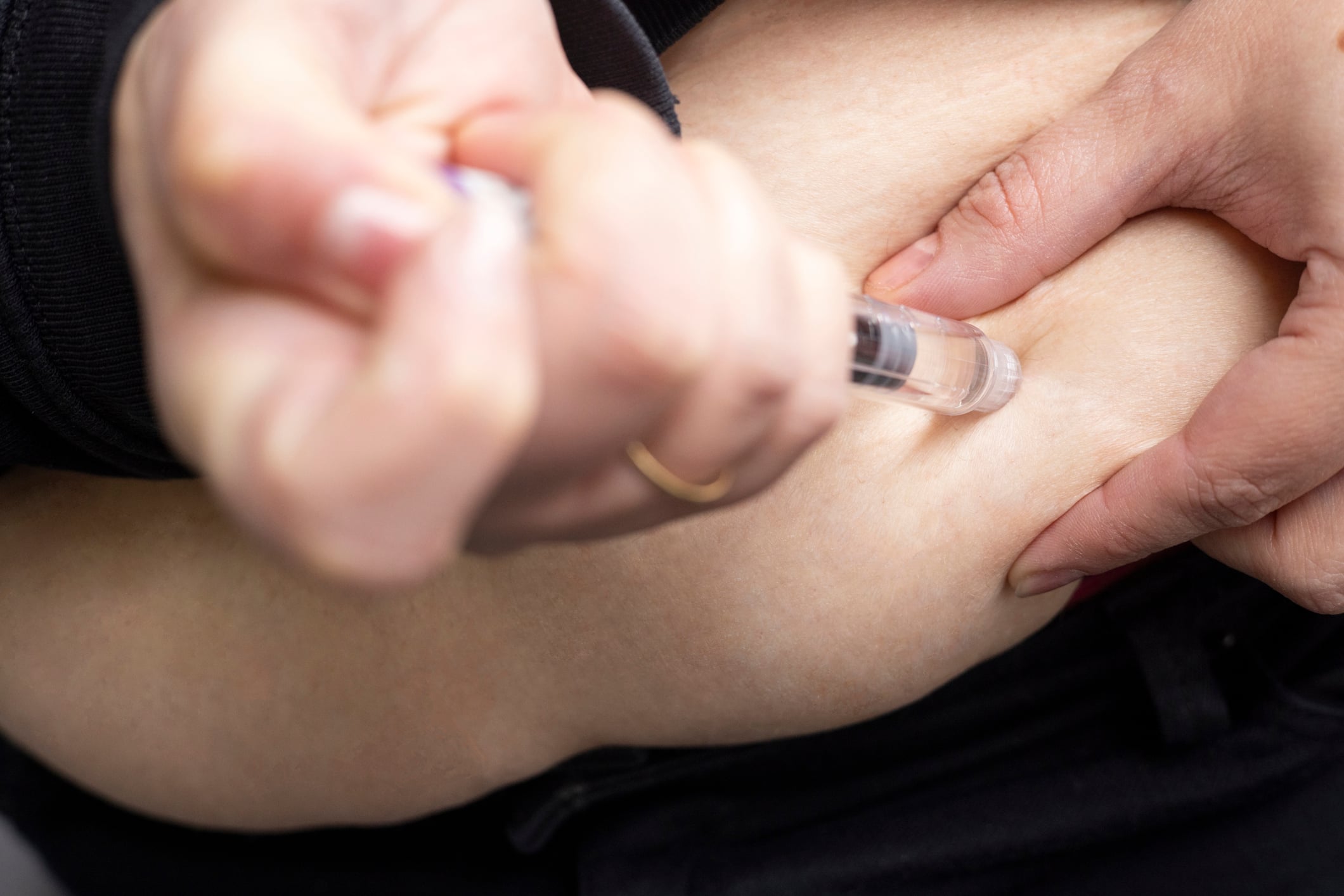
A treatment trial with omalizumab was launched for antihistamine-resistant light urticaria (n=13; 31.7%) [1]. Treatment with the anti-IgE antibody was introduced in 5 patients between 2015 and 2018, in 5 patients in 2019 and in 3 patients between 2020 and 2021. In this group (n=13), the median duration of omalizumab treatment was 51.4 months. The median basal UCT value was 6. Omalizumab was initially used in all patients at a dose of 300 mg every 4 weeks. Drug survival after 12, 24 and 48 months was 100% (13/13), 100% (12/12) and 88.9% (8/9) respectively. A dose increase of omalizumab to 450 mg every 4 weeks was required in 3 patients in whom partial/full control was subsequently achieved. No patient required an increase in dosage to 600 mg every 4 weeks. A gradual dose reduction was attempted in 8 patients, but only 5 patients were able to discontinue omalizumab completely. In four of these 5 patients, relapses of light urticaria occurred after discontinuation. A total of 4 patients will continue to be treated with 300 mg every 6 weeks and 3 patients with 300 mg every 8 weeks. Antihistamines were only used in 5/13 patients at the approved dose (1/5), 2-fold (3/5) or 3-fold (1/5) after 6 months.

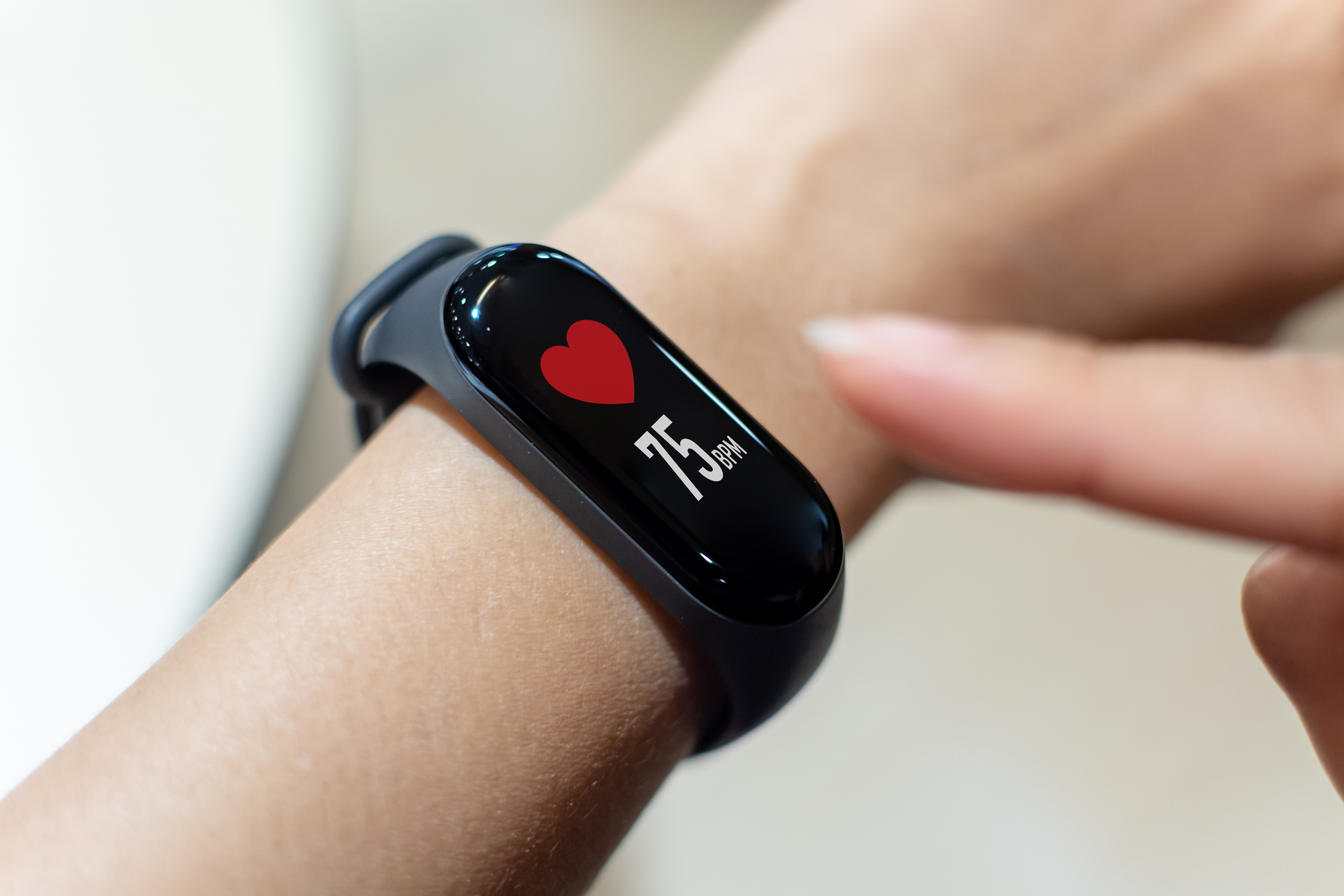
After starting treatment with omalizumab, UCT and FcεRI measurements were performed in 11 patients at weeks 4, 8, 12 and 24. A rapid and significant increase in median UCT values was observed, indicating an improvement in symptom control in patients treated with omalizumab. In addition, the change in FcεRI expression in basophils after the start of omalizumab treatment in the same period showed a significant decrease from baseline at week 4, which persisted until week 24. No adverse effects of omalizumab were observed in any patient.
Discussion
The demographic characteristics of the cohort are consistent with previous studies [6–8] and confirm that light urticaria is most common in young adult women. Regarding comorbidities, this study confirmed that atopic dermatitis and other CindU are more common in light urticaria. The clinical features of light urticaria and the results of photoprovocation were similar to those of previous studies. As far as laboratory diagnostic features are concerned, the absence of an autoimmune and/or inflammatory serologic background has also been postulated for other types of chronic urticaria, such as autoimmune CSU [9].
In the case of light urticaria, the exact determination of the range of triggering wavelengths is used to select sunscreens or light sources with an appropriate UV filter. However, it has been shown that in many patients the threshold for the relevant physical trigger is low and therefore complete avoidance of symptoms is practically impossible [12]. In individual cases, a three-day rush therapy with UV-A has proven to be effective in inducing tolerance [13–15]. As the tolerance induction only lasts for a few days, consistent daily exposure to the stimulus at the current threshold level is required. Second-generation H1 antihistamines (H1-AH-2G) are the symptomatic treatment of first choice. The combination of light-hardening and antihistamines has also achieved long-lasting therapeutic effects in some cases [13–15]. For patients who do not show a sufficient response to H1-AH-2G, omalizumab is the next step in the treatment algorithm. The recommended starting dose is 300 mg every 4 weeks, the dosage is independent of the total serum level.
The therapeutic characteristics in the present study indicate that light urticaria symptoms were well controlled in a significant proportion of patients with 2 times the dose of antihistamines, without the need for further therapeutic measures during follow-up [1]. After a median follow-up period of 60 months, a sustained complete remission was observed in around one third of patients. In addition, a low basal value in the urticaria control test (UCT; score for evaluating disease severity) was a significant clinical differential feature (p<0.01) for the administration of omalizumab. The study authors point out that this is a possible predictor for a lack of response to antihistamines and the subsequent prescription of omalizumab as well as for longer-lasting urticaria [1].
Previous publications have shown that light urticaria symptoms are triggered by the involvement of an IgE signaling pathway [3,4,10,11]. The association of clinical improvement with the reduction of FcεRI in omalizumab responders underlines the importance of the pathophysiology of IgE-mediated type 1 hypersensitivity in light urticaria in clinical practice. According to current knowledge, it is assumed that incident light is absorbed by a precursor molecule in the dermis, which leads to activation of the chromophore [2]. The resulting photoproduct acts as a photoallergen against which specific IgE is formed, which binds to mast cells in the skin. Attachment of the photoallergen to the FcεRI receptor of the IgE leads to an antigen-antibody reaction with subsequent mast cell degranulation, which results in the clinical symptoms of light urticaria with wheal formation, erythema and itching.
Literature:
- Pesqué D, et al.: Solar Urticaria: An Ambispective Study in a Long-term Follow-up Cohort with Emphasis on Therapeutic Predictors and Outcomes. Acta Derm Venereol 2024 Jan 8;104: adv25576.
- Gaebelein-Wissing N, et al.: Lichturtikaria: Klinik, Diagnostik, Verlauf und Therapiemanagement bei 27 Patienten. JDDG 2020;18(11): 1261–1269.
- Goetze S, Elsner P: Solar urticaria. JDDG Ges 2015; 13: 1250–1253.
- McSweeney SM, et al.: Pathogenesis of solar urticaria: Classic perspectives and emerging concepts. Exp Dermatol 2022; 31: 586–593.
- Haylett AK, et al.: Solar urticaria in 145 patients: assessment of action spectra and impact on quality of life in adults and children. Photodermatol, Photoimmunol, Photomedicine 2018; 34: 262–268.
- Morgado-Carrasco D, et al.: Clinical and photobiological response in eight patients with solar urticaria under treatment with omalizumab, and review of the literature. Photodermatol Photoimmunol Photomed 2018; 34: 194–199.
- Snast I, et al.: Real-life experience in the treatment of solar urticaria: retrospective cohort study. Clin Exp Dermatol 2019; 44: e164–e170.
- Photiou L, Foley P, Ross G. Solar urticaria – an Australian case series of 83 patients. Australas J Dermatol 2019; 60: 110–117.
- Pesqué D, et al.: Autoimmune diseases and low baseline immunoglobulin E in chronic spontaneous urticaria: a clinical and therapeutic prospective analysis in real clinical practice. J Allergy Clin Immunol Pract 2023; 11: 3763–3771.e5
- McSweeney SM, et al.: Systematic review of the clinical characteristics and natural history of solar urticaria. JAAD 2023; 89: 138–140.
- Maurer M, et al.: Management and treatment of chronic urticaria (CU). JEADV 2015; 29: 16–32.
- Lehmann P, Schwarz T: Lichtdermatosen: Diagnostik und Therapie. Dt Arztebl Int 2011; 108(9): 135–141.
- Zuberbier T, et al.: Deutsche S3-Leitlinie zur Klassifikation, Diagnostik und Therapie der Urtikaria, 2022. AWMF-Leitlinienregister (013-028).
- Beissert S, et al.: UVA rush hardening for the treatment of solar urticaria. Case Reports. JAAD 2000; 42(6): 1030–1032.
- Hölzle E: Lichturtikaria. In: E Hölzle: Photodermatosen und Lichtreaktionen der Haut. Wissenschaftliche Verlagsgesellschaft, Stuttgart, 2003: 130–153.
DERMATOLOGIE PRAXIS 2024; 34(1): 26–27