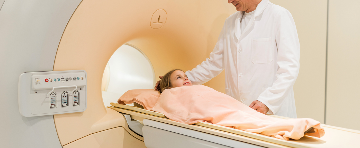
Regular examination of the lungs using magnetic resonance imaging (MRI) can record how lung disease develops in children with CF. In addition, if the disease is detected and treated during newborn screening (NGS) before symptoms appear, it may be milder. This is what scientists from the German Center for Lung Research (DZL) found out in a study that followed preschool children for four years.
“The MRI scans showed that changes in the lungs already increase over the first four years of life, such as the bronchial walls thickening and increasingly certain areas of the lungs not being as well ventilated because the airways are blocked with mucus,” says private lecturer Dr. Mirjam Stahl, a DZL researcher at Charité – Universitätsmedizin Berlin. In her study, she used MRI once a year on nearly 100 children up to four years old. All children had cystic fibrosis detected either by newborn screening or on the basis of clinical symptoms typical of cystic fibrosis. Cystic fibrosis is a congenital multi-organ disease in which a salt transport disorder produces viscous secretions that gradually impair the function of many organs. The disease is caused by defects in a gene that contains the blueprint for the CFTR chloride channel. The disease cannot be cured. One way to treat the symptoms is for the children to inhale bronchodilators and expectorants.
Start of therapy in the symptom-free stage brings advantages
The study also found that changes in children’s lungs were not as pronounced if their disease was detected through newborn screening and treatment was started before the first symptoms appeared. “Starting therapy early in the asymptomatic stage keeps the disease at a lower level but cannot stop its progression,” Stahl said. She hopes that a new group of drugs that directly target the defective chloride channel, known as CFTR modulators, could have an even greater effect here and possibly slow down the progression of the disease. Such questions would have to be investigated by clinical studies in the future.
Uniform specifications are lacking
Since 2016, newborn screening has included testing for the presence of cystic fibrosis. First, the babies’ blood is analyzed. If certain values are conspicuous, a sweat test is performed to confirm the diagnosis. To assess whether the lungs are already affected, doctors may use pulmonary function tests or imaging techniques such as a lung X-ray, MRI or computed tomography (CT) scan. MRI and CT have better spatial resolution than X-ray and therefore reveal more detail. MRI offers the advantage over CT that it is radiation-free. However, which procedure is used depends on the clinic, as there are no uniform guidelines for this to date.
Source:
Magnetic Resonance Imaging Detects Progression of Lung Disease and Impact of Newborn Screening in Preschool Children with Cystic Fibrosis. Stahl M, Steinke E, Graeber SY, Joachim C, Seitz C, Kauczor HU, Eichinger M, Hämmerling S, Sommerburg O, Wielpütz MO, Mall MA.Am J Respir Crit Care Med. 2021 Jul 20.
DOI: 10.1164/rccm.202102-0278OC
https://www.atsjournals.org/doi/10.1164/rccm.202102-0278OC