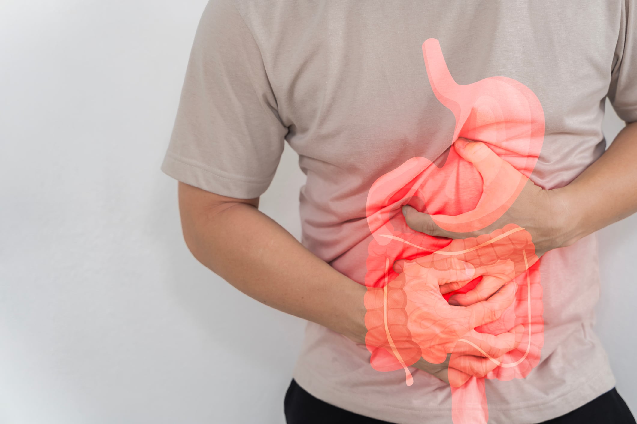
Abdominal pain in the right lower abdomen, accompanied by nausea or fever, is a typical symptom of appendicitis. If there is a classic symptomatology for appendicitis, the diagnosis can be made clinically. However, there are also atypical symptoms and courses, particularly in young children, older patients and pregnant women. In patients with atypical or doubtful findings, however, imaging procedures should be used promptly.
Acute appendicitis is the most common cause of surgical intervention for acute abdomen. Over 5% of the population will develop appendicitis at some point in their lives. It is most common in the teenage years and in the third decade of life, but can occur at any age [3]. In children and adolescents, several differential diagnoses [6] must be considered (Table 1). Other findings that can occur under the picture of appendicitis are carcinoids, carcinomas, villous adenomas and diverticula. The appendix can also be affected in Crohn’s disease and ulcerative colitis with pancolitis (inflammatory bowel disease).

In most cases, the cause of appendicitis is an obstruction of the appendix lumen, typically caused by lymphoid hyperplasia, more rarely by fecal stones, foreign bodies or even parasites. The occlusion leads to inflammatory edema, bacterial overgrowth, ischemia and inflammation. Typical pathogens of appendicitis include Escherichia coli, Pseudomonas aeruginosa or Klebsiella pneumoniae [1]. If left untreated, necrosis, gangrene and perforation occur. If the perforation is enveloped by the surrounding mesh, an abscess may form. Overview 1 lists the symptoms and clinical signs of appendicitis.

The different stages of appendicitis are listed in Overview 2.

If symptoms persist postoperatively, depending on the stage of appendicitis, a postoperative complication with abscess or pronounced inflammatory reaction with hypertrophic scarring must also be considered, as shown in case study 2 .
X-ray examinations are of no value in the diagnosis of acute appendicitis.
In recent years, sonography has established itself as a reliable tool in the diagnosis of appendicitis. Meta-analyses attest the method a sensitivity of up to 87% and a specificity of up to 94%. Abdominal ultrasound typically shows an enlarged appendix diameter of more than 6 mm, wall thickening and a cockade phenomenon as well as a circular accumulation of fluid around the organ. Hyperemia can often be detected by color Doppler.
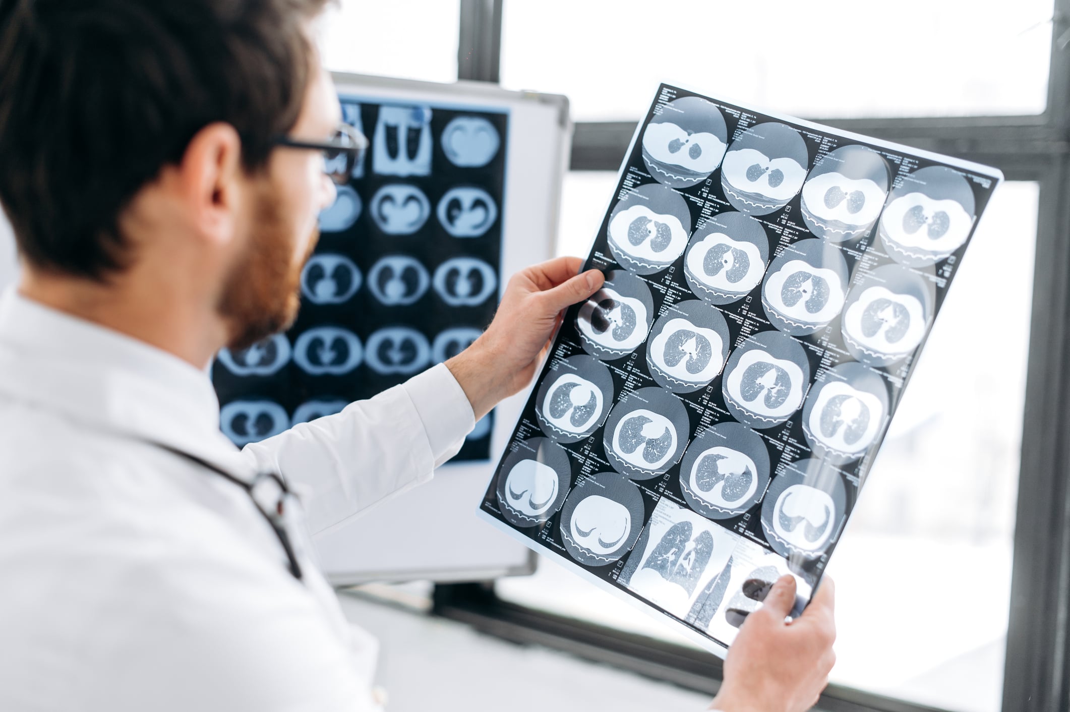
The use of imaging is now standard in the diagnosis of appendicitis. This reduces the number of negative surgical interventions and the overall treatment costs. Although ultrasound is often used in pediatric patients to avoid radiation exposure, a CT scan is the most accurate way to make a diagnosis in adults, according to a report in the British Journal of Surgery [2]. The accuracy of the scans in identifying patients with and without acute appendicitis was 98 and 98.5 percent respectively.
Magnetic resonance imaging can be used as an alternative to CT in children and adolescents in order to avoid radiation exposure [4]. Signs of appendicitis in the contrast-enhanced MRI examination were fluid accumulation with wall thickening, atypical contrast enhancement in the bowel wall, appendix enlargement (>6 mm), fecal stones (appendicoliths), inflammatory fat stranding around the appendix, and diffusion disorders. Accumulations of fluid in the immediate vicinity, phlegmon and abscesses accumulating at the edges even without a recognizably inflamed appendix were considered signs of appendix perforation [5].
Case study
In case example 1 (Fig. 1A to 1C), MRI shows acute appendicitis in a 22-year-old female patient. The appendix showed an inflammatory edematous wall thickening to 5 mm and a diameter of 1.3 cm. An inflammatory environmental reaction in the fatty tissue was also already recognizable in the KM sequences. The symptoms were recurrent for four weeks and the inflammatory parameters were alternately slightly elevated. The operation performed immediately after the MRI confirmed the acute, preperforating appendicitis.

Case 2 demonstrates (Fig. 2A to 2C) the condition after perforated appendicitis, local abscess and peritonitis. Wound healing disorder with local phlegmon. In the 18-year-old patient, a pronounced scar reaction with persistent pain was detected two months after the operation and a recurrent abscess was ruled out.

Take-Home-Messages
- Appendicitis often presents with typical symptoms in the right lower abdomen, but can also have variable symptoms and laboratory values that deviate from the norm.
- In young patients in particular, several differential diagnoses must be considered.
- Imaging examinations are very important for assessing the stage of the inflammation in order to avoid a life-threatening escalation.
- Surgical treatment leads to definitive rehabilitation and avoids complications.
Literature:
- «Appendizitis», https://flexicon.doccheck.com/de/Appendizitis, (last accessed 19.03.2024)
- Haijanen J, et al.: Diagnostic accuracy using low-dose versus standard radiation dose CT in suspected acute appendicitis: prospective cohort study. Br J Surg 2021; 108(12): 1483–1490.
- «Gastrointestinale Erkrankungen», www.msdmanuals.com/de-de/profi/gastrointestinale-erkrankungen, (last accessed 19.03.2024)
- Dingemann J, Ure B: Imaging and the Use of Scores for the Diagnosis of Appendicitis in Children. Eur J Pediatr Surg 2012; 22(03): 195–200.
- Koning JL, Naheedy JH, Kruk PG: Diagnostic performance of contrast-enhanced MR for acute appendicitis and alternative causes of abdominal pain in children. Pediatr Radiol 2014; 44: 948–955.
- Staatz G, Schneider K: Differenzialdiagnose des akuten Abdomens. Teil IV (akutes Abdomen im Kindesalter). Radiologie up2date 2; 2010: 103–116.
- Wiesner W, Kirchhoff TD, Opherk JP: Differenzialdiagnose des akuten Abdomens. Teil II. Radiologie up2date 1; 2009; 35–45.
HAUSARZT PRAXIS 2024; 19(4): 47–49